

Hope this scan is something quick you can add on to your current scanning repertoire.
Case presentation
Presentation
You are working in Crit and EMS rolls a gurney in with a 58-year-old female as a stroke code.
Ninety minutes ago, while folding laundry and talking to her sister on speaker, she started having difficulty with the clothes. She noticed her left arm felt heavier and she was uncoordinated folding the shirts. She felt clumsy and mentioned it to her sister, who became concerned that she might be having a stroke and told her to call 911. The patient said maybe she had just overworked her arm vacuuming earlier that day and that she was going to lie down to rest.
About 45 minutes later, her sister called back to check on her and noticed that the patient sounded strange on the phone, like she had a sore throat. Her arm still felt heavy, and her sister insisted that she call 911.
History
PMH: hypertension and hyperlipidemia; sometimes forgets to take her medications.
Social: former smoker (quit about 8 years ago); drinks alcohol socially.
Denies: headache or head trauma, dizziness, visual disturbances, recent illness, sore throat, URI symptoms, chest pain, palpitations, shortness of breath, abdominal pain, nausea, vomiting, diarrhea.
Vital signs
HR 94 · BP 184/96 · RR 18 · Temp 98.8 · SpO₂ 96% RA · FSG 185
Physical exam
General: no acute distress, speaking in a raspy whisper.
HEENT/Neuro: normocephalic, atraumatic. Right pupil 4 mm; mild left ptosis with a 2 mm pupil. No other facial droop.
Strength: RUE 5/5, LUE 3/5, RLE 5/5, LLE 5/5. Sensation normal in all extremities.
Chest: regular rate and rhythm, lungs clear bilaterally.
Abdomen: nontender, nondistended.
ED course
You order the pertinent stroke brain imaging, and the patient rolls to CT, where the Stroke PA meets her in the hallway and completes the NIHSS: 3.
About 5 minutes after returning from CT, she starts to feel some mild fluttering in her chest that is going into her neck, along with some chest discomfort.
EKG: NSR, HR 99, no ischemic changes.
Chest X-ray: normal.
Troponin: cooking with the rest of the labs.
Still waiting on CT results and neurology recommendations.
What can you do now? POCUS!
You grab the ultrasound and start your bedside echo. The standard views all look normal: parasternal long, parasternal short, apical 4-chamber, and subxiphoid. You decide to add another view: the SSN.
The suprasternal notch (SSN) view
No, it’s not your social security number. It’s the suprasternal notch view! Before we get to the punchline, let’s learn or review it.
Adding the SSN view to your bedside echo extends your visual field to the distal ascending aorta, the transverse aortic arch, the major brachiocephalic vessels, and the proximal descending thoracic aorta.
Image acquisition
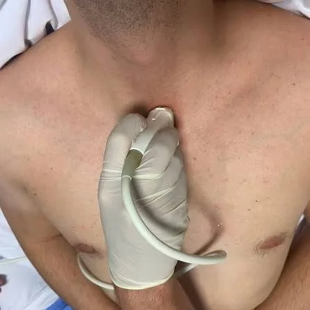
Position the patient: supine with the neck mildly hyperextended (you may use a rolled towel between the shoulders). It may also help to have the patient turn their head to the right.
Choose the probe: cardiac (phased-array).
Place it in the sternal notch vertically, with the indicator at the 12 o’clock position.
Rotate slightly so the indicator is at the 1 to 2 o’clock position.
Angle toward the heart if needed.
Anatomy
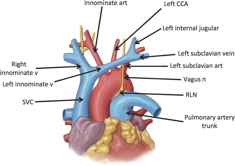
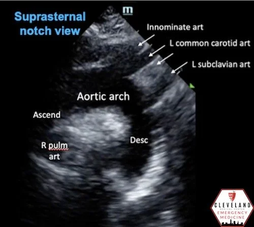
When aligned correctly in the long-axis plane of the arch, the anatomy forms a distinct curve often called the “candy cane” view:
The aortic arch: rests slightly oblique. The ascending aorta starts on the left, traverses into the arch, and ends with the descending aorta on the right.
The “three chimneys” (brachiocephalic branches), arising superiorly from the convexity of the arch:
Brachiocephalic (innominate) artery: most proximal, largest branch.
Left common carotid artery: middle branch.
Left subclavian artery: most distal branch before the aorta transitions to the descending segment.
Right pulmonary artery (RPA): seen in cross-section as an anechoic circle immediately underneath the arch.
Left atrium (LA): inferior to the RPA.
Normal aortic arch
The labeled still is on the left, and Riana’s aortic arch on video is on the right. Shout out to Riana’s aortic arch!
Back to the patient
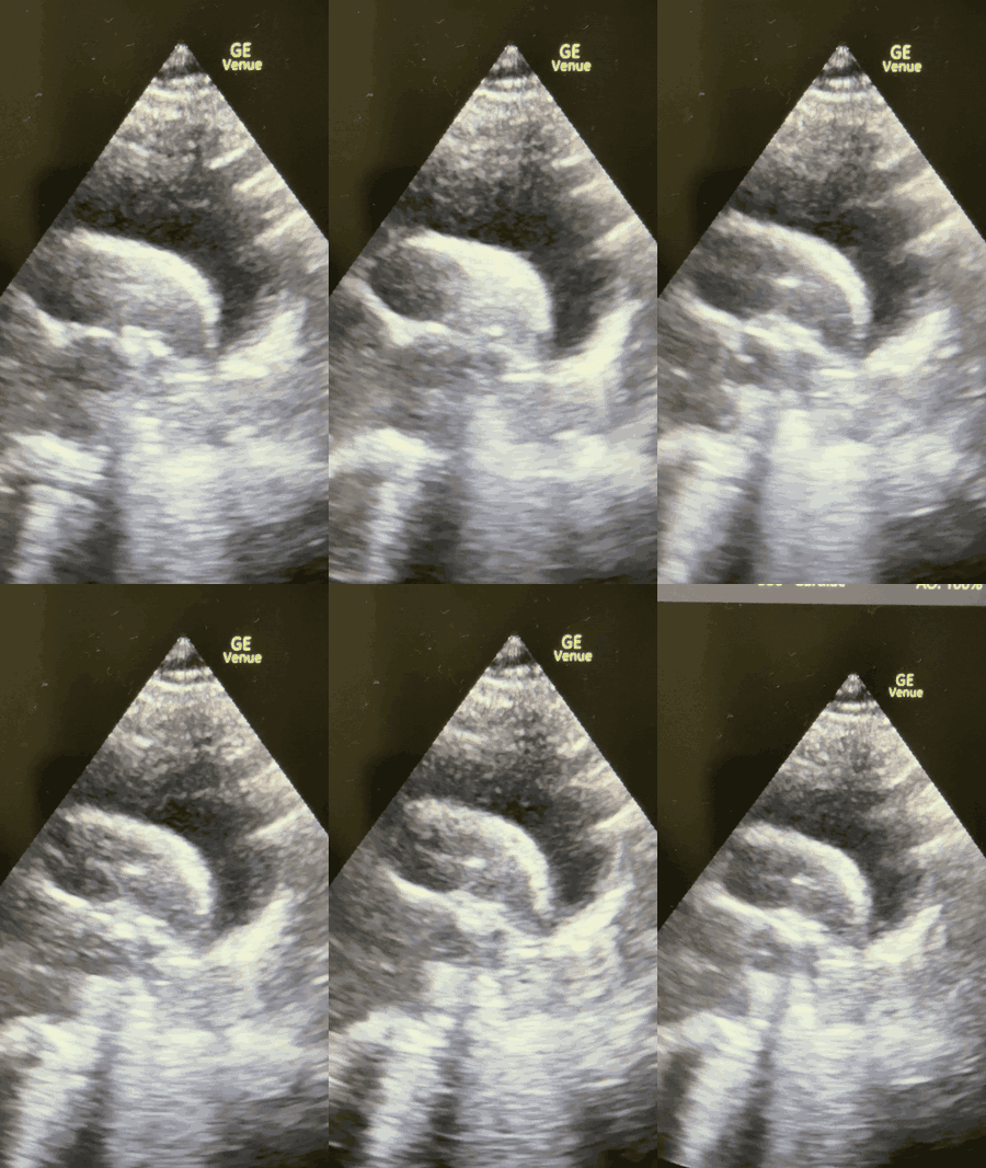
Unfortunately for your patient, her SSN view looked like this:
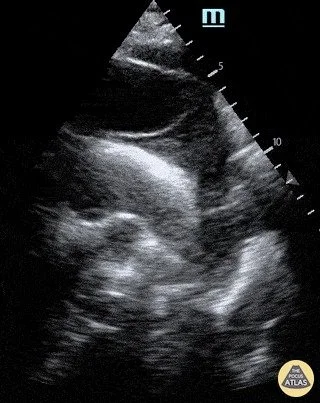
Diagnosis: type A ascending aortic dissection!
Meanwhile, Neurology sends you an Epic chat message:
“All imaging negative. Place in OBS for MRI brain.”
Image sources
The POCUS Atlas (thepocusatlas.com)
Prague ICU (pragueicu.com)
Anesthesia Key (aneskey.com)