

A systematic approach to AV blocks
Bradydysrhythmias can be confusing without a step-by-step approach, especially when non-conducted beats are involved. Three bradydysrhythmias with dropped QRS complexes need careful evaluation and pattern recognition to tell apart:
1. Second-degree AV block type I (Mobitz I/Wenckebach)
2. Second-degree AV block type II (Mobitz II)
3. Third-degree AV block (complete heart block)
Step 1: Map out every P wave
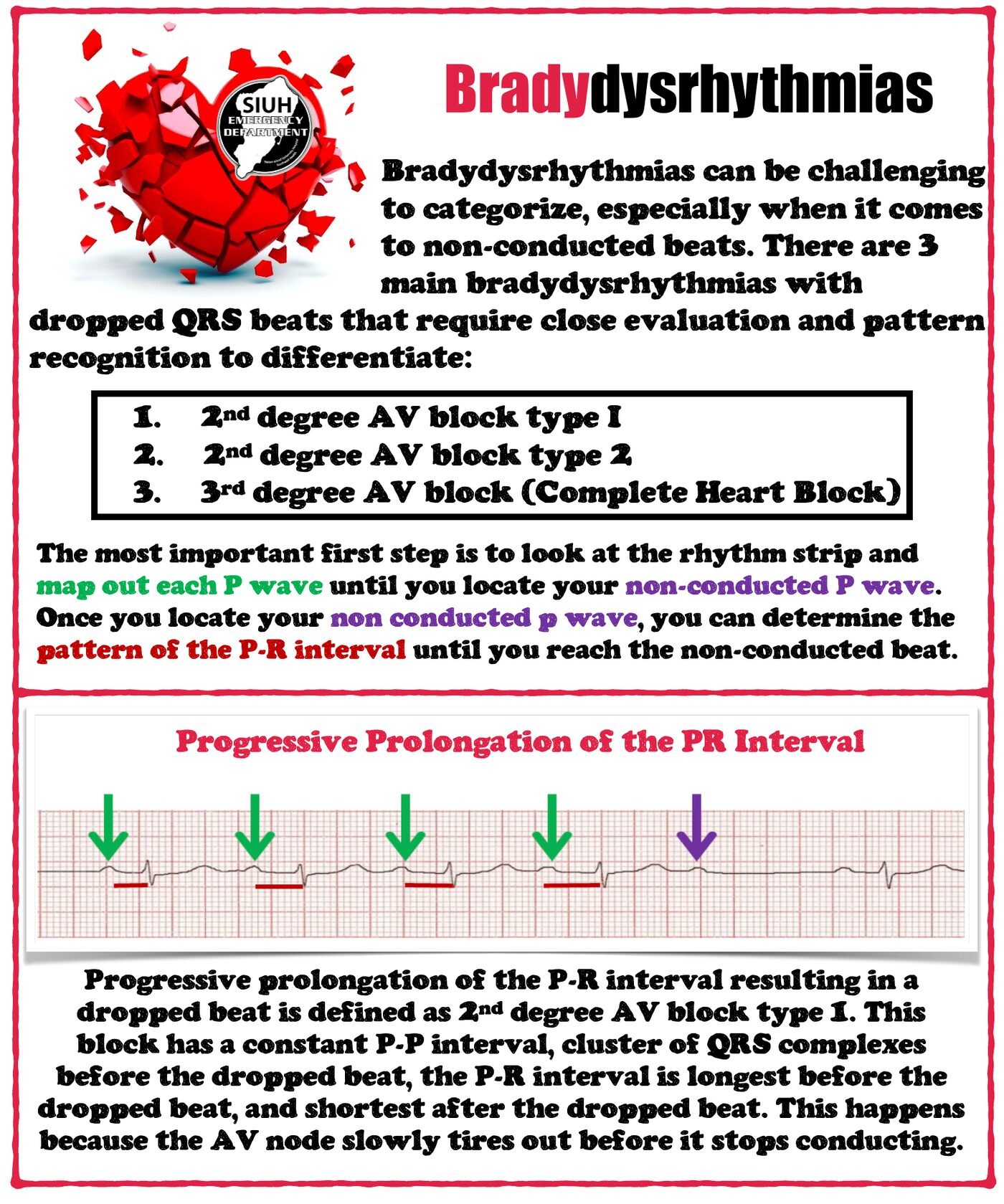
Look at the rhythm strip and mark each P wave until you find the non-conducted P wave. Then look at how the PR interval behaves in the beats leading up to the dropped beat.
The secret to AV blocks is the PR interval.
Step 2: Read the PR interval pattern
Progressive prolongation → second-degree AV block type I
The PR interval lengthens progressively until a beat is dropped.
Constant P–P interval.
QRS complexes cluster ("group beating") before the dropped beat.
The PR interval is longest just before the dropped beat and shortest just after it.
Mechanism: the AV node gradually tires out until it fails to conduct.
Constant PR → second-degree AV block type II
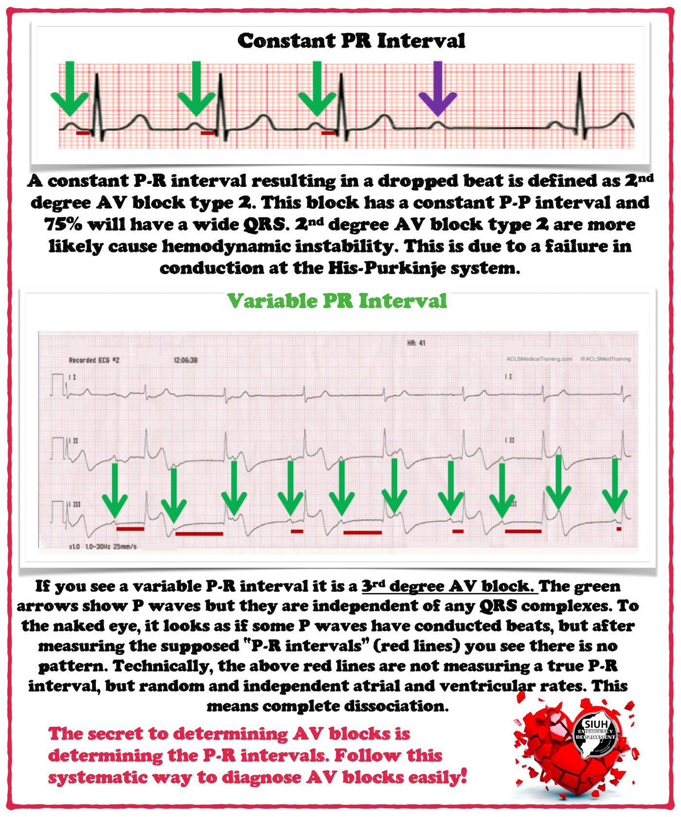
A constant PR interval with a suddenly dropped beat.
Constant P–P interval.
About 75% have a wide QRS.
More likely to cause hemodynamic instability, because the conduction failure is in the His–Purkinje system.
Variable PR → third-degree AV block
P waves and QRS complexes are independent: complete AV dissociation.
Some P waves may appear to conduct, but measuring the apparent PR intervals reveals no pattern. These are not true PR intervals, just random alignment of independent atrial and ventricular rates.
Quick reference
| PR interval before the dropped beat | Diagnosis | Level of block |
|---|---|---|
| Progressively lengthens | 2nd-degree type I (Wenckebach) | AV node |
| Constant | 2nd-degree type II (Mobitz II) | His–Purkinje (often wide QRS) |
| Variable/no relationship | 3rd-degree (complete heart block) | AV dissociation |
Clinical pearls
Find the P waves first. Calipers or a marked-up strip make the pattern obvious.
Wenckebach is usually nodal and often benign, but always interpret it in the clinical context.