

Back to basics: the four core cardiac ultrasound views
Continuing a cardiology theme, this edition returns to the basics of bedside cardiac ultrasound: how to get each standard view and what to look for in each.
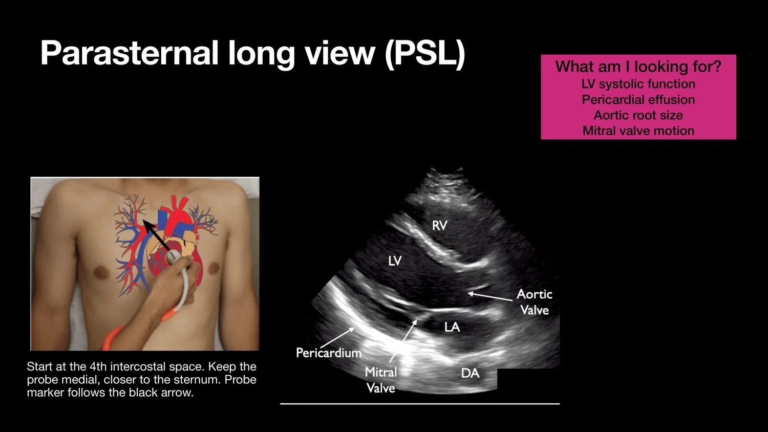
Parasternal long axis (PSL)
Probe position: start at the 4th intercostal space, keeping the probe medial, close to the sternum. The marker orientation is shown in the PDF.
What to look for:
LV systolic function
Pericardial effusion
Aortic root size
Mitral valve motion
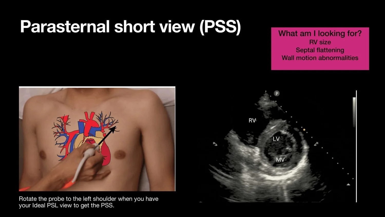
Parasternal short axis (PSS)
Probe position: from an ideal PSL view, rotate the probe toward the patient's left shoulder.
What to look for:
RV size
Septal flattening (the "D-sign")
Wall motion abnormalities
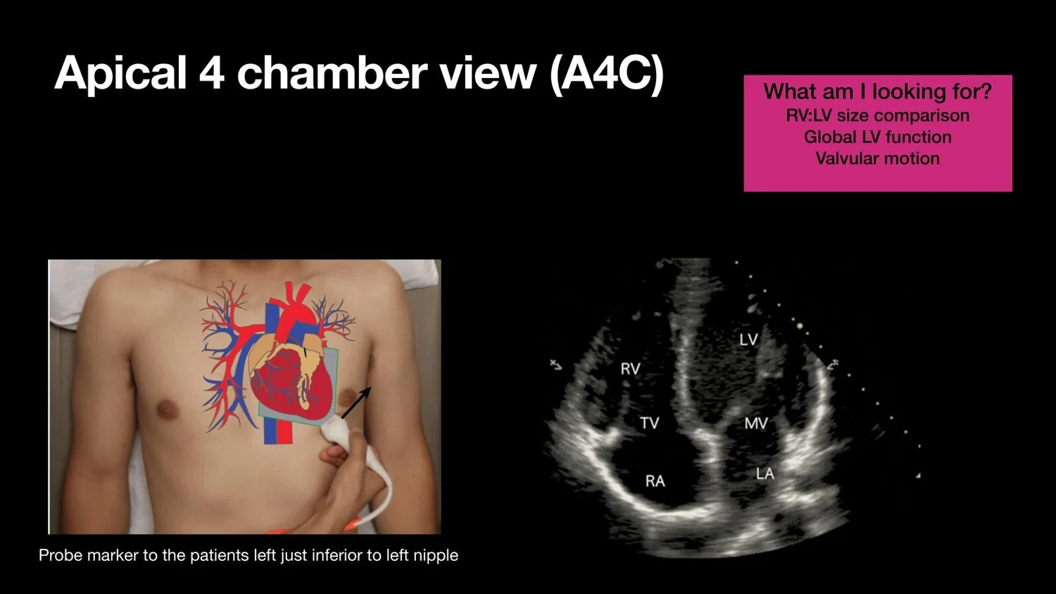
Apical 4-chamber (A4C)
Probe position: marker toward the patient's left, just inferior to the left nipple.
What to look for:
RV:LV size comparison
Global LV function
Valvular motion
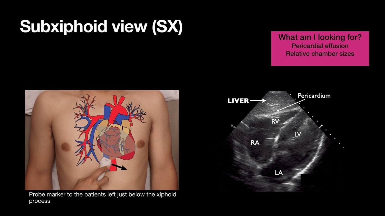
Subxiphoid (SX)
Probe position: marker toward the patient's left, just below the xiphoid process.
What to look for:
Pericardial effusion
Relative chamber sizes
Quick reference
| View | Where | Key questions |
|---|---|---|
| PSL | 4th ICS, close to the sternum | LV function, effusion, aortic root, mitral valve |
| PSS | Rotate from PSL toward left shoulder | RV size, septal flattening, wall motion |
| A4C | Below left nipple, marker to patient's left | RV:LV ratio, global LV function, valves |
| Subxiphoid | Below xiphoid, marker to patient's left | Effusion, relative chamber sizes |
Clinical pearls
No single view answers every question. Use at least two views to confirm a finding.