

Clinical scenario
A 5-year-old girl with idiopathic hydrocephalus status post VP shunt, and tonic-clonic seizures on levetiracetam 1 g daily, is brought in by her father for acting off her baseline for 2 days. He denies nausea or vomiting. She is lethargic, and you notice what appears to be a bilateral inferior gaze palsy.
What do you do next?
What is a VP shunt?
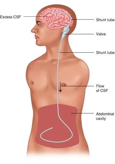
A ventriculoperitoneal (VP) shunt drains excess cerebrospinal fluid (CSF) from the ventricles to the peritoneum when normal CSF flow is obstructed, most commonly at the cerebral aqueduct connecting the third and fourth ventricles. The resulting pressure compresses brain tissue, and children present with seizures and altered mental status.
Anatomy of the shunt
A proximal catheter runs from the ventricle through a burr hole in the skull.
It drains into a reservoir with a one-way valve to prevent backflow. The reservoir can usually be palpated behind the right ear.
Distal tubing runs from the reservoir to the peritoneum. In children, extra tubing is coiled in the abdomen to allow for growth.
Complications
The most common are mechanical obstruction, fracture, and infection.
Obstruction: signs of increased ICP, including headache, seizures, and gaze palsies.
Infection: meningeal signs such as fever, headache, neck pain, and altered mental status.
History
In a child with a shunt whose parent reports altered mental status from baseline, assume shunt failure until proven otherwise.
Physical exam
Look for signs of increased ICP:
Cushing's triad: widened pulse pressure/hypertension, bradycardia, and irregular breathing
Pupillary reactivity
Gaze palsy ("sunset eyes")
Palpate the reservoir behind the ear and note whether it is compressible.
Work-up
If febrile, perform a standard sepsis work-up.
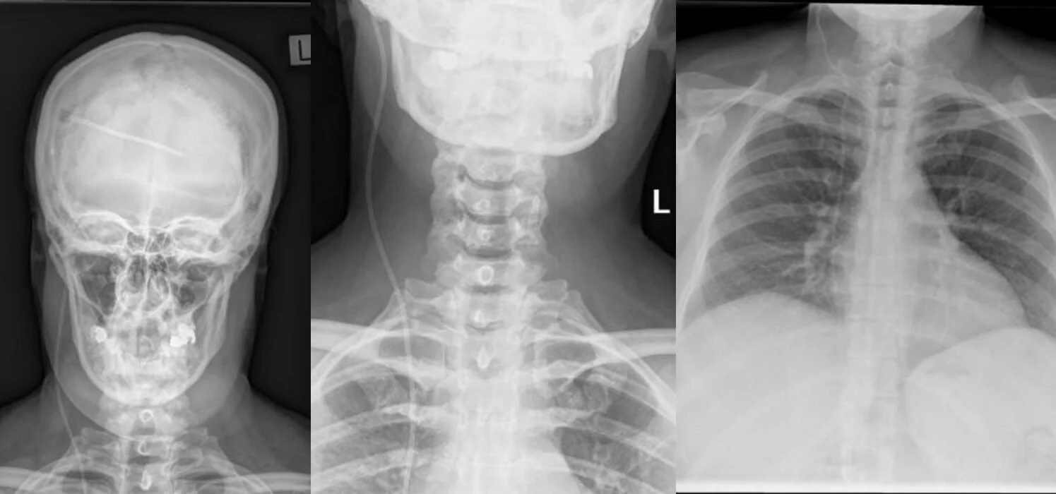
Shunt series for every patient with suspected shunt complication: four X-rays (skull, cervical spine, chest, and abdomen) to look for kinking or fracture along the tubing.
Non-contrast CT head if obstruction is suspected, to assess ventricular size.
Normal-appearing ventricles do not exclude shunt failure. If suspicion is high, neurosurgery may still tap the shunt.
Interventions and disposition
Stable patients
Infected shunts: admit for IV antibiotics and shunt revision.
Obstructed shunts: admit for revision.
Unstable or peri-arrest patients
Elevate the head of the bed, intubate, and prepare to tap the shunt.
The tap should preferably be done by neurosurgery. In dire circumstances, an emergency physician can do it in conjunction with neurosurgery.
Shunt tap technique
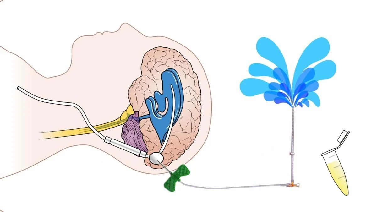
Attach a butterfly needle to tubing with a three-way stopcock and manometer.
Insert the needle into the reservoir (around the right ear).
Allow CSF to drain passively; do not aspirate. Continue until vital signs improve. Often a small amount, such as 10 mL, can reduce the risk of herniation.
Obstruction is often proximal to the reservoir, most commonly when the proximal catheter tip is caught in the choroid plexus. In that case the tap may fail, and emergent neurosurgery is life-saving.
Clinical pearls
Shunt + altered mental status = shunt failure until proven otherwise.
Sunset eyes, bradycardia, and hypertension are late, ominous signs of raised ICP.
A normal CT does not clear the shunt. Call neurosurgery when suspicion is high.
Shunt tap: drain, don't aspirate.