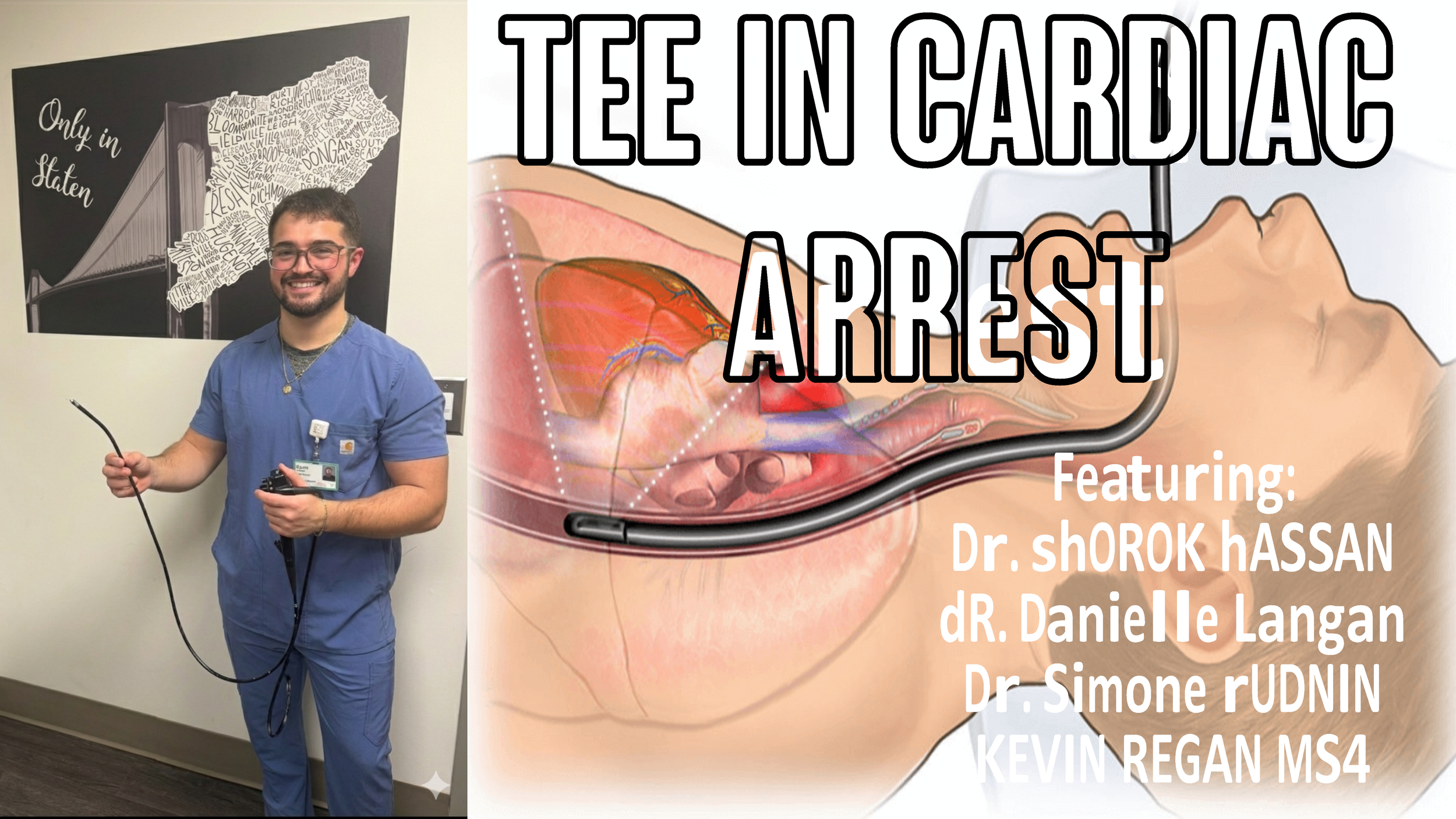
Episode 35: TEE in Cardiac Arrest
Learning Points
TEE in Cardiac Arrest
Featuring Dr. Shorok Hassan, Dr. Danielle Langan, Dr. Simone Rudnin & Kevin Regan MS4
1. External CPR landmarks can be imprecise. TEE shows, in real time, whether the compressor’s hands are over the LV or unintentionally over the LVOT, allowing immediate correction that can meaningfully improve perfusion.
2. Unlike TTE, which requires interrupting CPR and competing for space on the chest wall, TEE provides uninterrupted visualization from inside the esophagus. This helps minimize pauses both during compressions and pulse checks.
3. With TEE, clinicians can directly watch LV filling, ejection, valve motion, and even the quality of each compression. Additionally we can visualize other findings such as tension pneumothorax, hypovolemia, and fine V-fib.
4. The use of TEE does have a learning curve, and can be cost prohibitive. Also, many EDs don’t yet have credentialed physicians, which limits widespread adoption.
5. ACEP acknowledges the potential of TEE during cardiac arrest but highlights the lack of large, outcomes-driven studies demonstrating improved survival or neurological function. Most existing data comes from small, observational studies reflecting how hard it is to conduct high quality research during chaotic, time-critical resuscitations.