Critical Care Casebook Episode 5: Central retinal artery occlusion
Presented by Dr. Eli Azrak, Dr. Brendan Freeman, Dr. Sarah Pokrzywa.
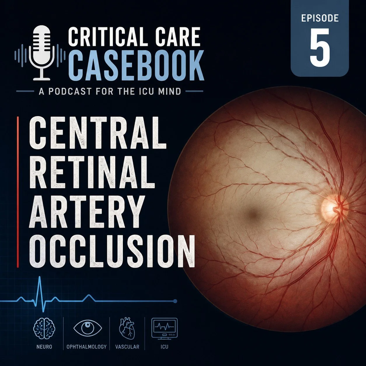
A 78-year-old woman presents with sudden, painless monocular vision loss and is ultimately diagnosed with a central retinal artery occlusion (CRAO), an ocular stroke. The case explores the emergency evaluation of acute vision loss and highlights several practical management lessons.
What causes painless monocular vision loss?
The differential includes CRAO, central retinal vein occlusion, retinal detachment, vitreous hemorrhage, and giant cell arteritis. In an older patient with abrupt, severe vision loss and an otherwise normal neurologic exam, CRAO should be considered until proven otherwise.
Should CRAO trigger a stroke code?
Yes. CRAO is considered a form of ischemic stroke. Patients require rapid neurologic assessment, brain imaging, vascular imaging when appropriate, and consultation with neurology and ophthalmology.
Can ocular ultrasound help?
Absolutely. Point-of-care ultrasound can rapidly identify retinal detachment, vitreous hemorrhage, and the retrobulbar spot sign—a bright echogenic focus at the optic nerve head representing an embolus within the central retinal artery. In this case, identification of the spot sign strongly supported the diagnosis before ophthalmologic confirmation.
How should we handle contrast refusal?
Rather than arguing with patients, explore the reason for refusal. Many patients fear kidney injury from contrast despite modern evidence showing a very low risk in patients with normal renal function. Shared decision-making and addressing specific concerns can often improve acceptance of recommended testing.
What about thrombolytics?
Historically, CRAO patients were often considered for IV thrombolysis if they presented within the treatment window. However, emerging evidence—including a recent randomized controlled trial published in the New England Journal of Medicine—found no improvement in visual outcomes with thrombolytics compared with aspirin, while exposing patients to greater bleeding risk, including intracranial hemorrhage.
So what should emergency physicians do now?
The initial workflow remains unchanged: activate the stroke pathway, obtain imaging, involve neurology and ophthalmology early, and evaluate for underlying embolic disease. What may be changing is the routine use of thrombolytics, as current evidence increasingly suggests that the risks outweigh the benefits for most CRAO patients.
Clinical Pearls
CRAO is an ocular stroke and should be treated as a time-sensitive emergency.
Severe vision loss may be profoundly disabling despite a low NIH Stroke Scale score.
Ocular POCUS can identify the retrobulbar spot sign and rapidly narrow the differential.
Modern evidence does not currently support routine thrombolytic therapy for CRAO.
Rapid diagnosis remains critical because these patients require urgent stroke evaluation and secondary prevention.