

Clinical scenario
A 68-year-old man with hypertension, hyperlipidemia, prior appendectomy, small bowel resection, and recurrent small bowel obstructions presents with 2 days of worsening abdominal pain, nausea, vomiting, and distension. His last bowel movement was yesterday, and he has not passed flatus since. He says it feels like his previous obstructions.
Vitals: BP 128/74, HR 104, RR 18, T 37.1 °C
Exam: distended, tympanitic abdomen with diffuse tenderness, worst around the umbilicus. No guarding, rebound, or rigidity.
You grab the ultrasound. What are you looking for? Small bowel obstruction (SBO).
How to scan for SBO
Probe: curvilinear.
Technique: start in the right lower quadrant and sweep systematically across the abdomen in a "lawnmower" pattern.
Image abnormal bowel in both transverse and longitudinal views.
Evaluate multiple loops for:
diameter
peristalsis
bowel-wall appearance
free fluid
What does normal small bowel look like?
Relatively small caliber
Active, coordinated peristalsis
Normal bowel-wall layers
Plicae circulares traversing the lumen
This patient's ultrasound
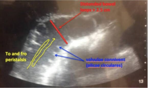
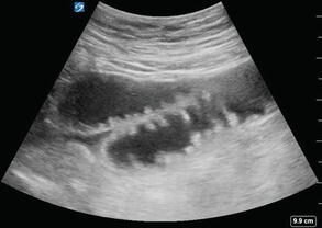
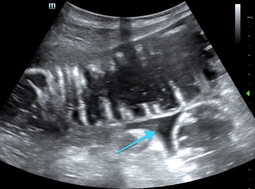
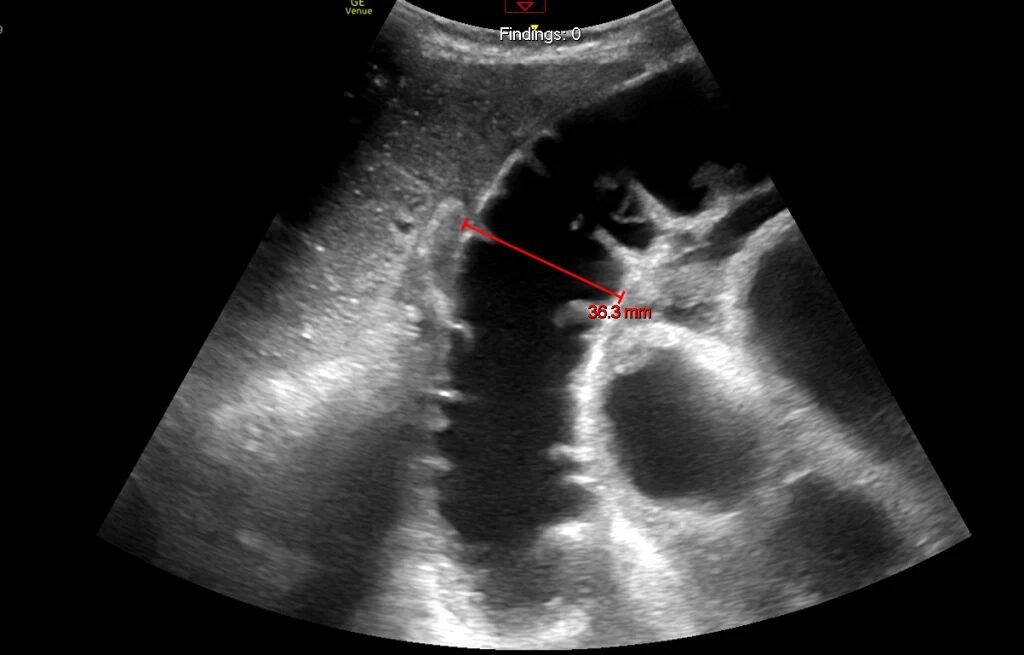
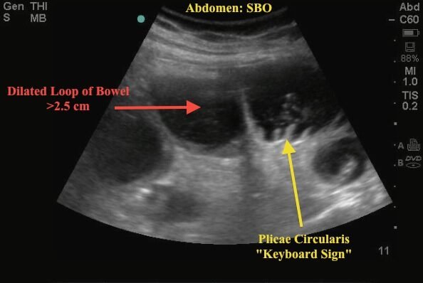
The images (Figures 1–4 and the linked PDF) show multiple dilated, fluid-filled small bowel loops with prominent plicae circulares, producing the characteristic "keyboard" or "piano key" appearance. There is also anechoic free fluid between the dilated loops, forming the triangular "tanga" sign.
Classic POCUS findings in SBO
| Finding | What you see |
|---|---|
| Dilated bowel | Small bowel > 2.5 cm in diameter is abnormal |
| Keyboard/piano-key sign | Prominent plicae circulares visible in dilated, fluid-filled bowel |
| To-and-fro sign | Contents shuttle back and forth instead of moving forward, because they cannot pass the obstruction |
| Tanga sign | Triangular (thong-shaped) free fluid between dilated loops |
Management
NPO
IV fluids
Analgesia
Antiemetics
Nasogastric decompression
General surgery consultation
Clinical pearls
Dilated (> 2.5 cm) fluid-filled loops plus to-and-fro peristalsis at the bedside: think SBO and call surgery early.
POCUS can speed the diagnosis while you arrange definitive imaging and decompression.