
Ultrasound differentiation of epididymitis, orchitis, and torsion
An image-based review of acute scrotal pain: the buddy shot, increased flow in epididymitis/orchitis, reactive hydrocele, testicular torsion with return of flow after detorsion, and a necrotic testicle.
Clinical image review: acute scrotal pain
For a patient with acute scrotal pain, compare the appearance of the testes and the Doppler flow patterns in the examples below (see the image gallery).
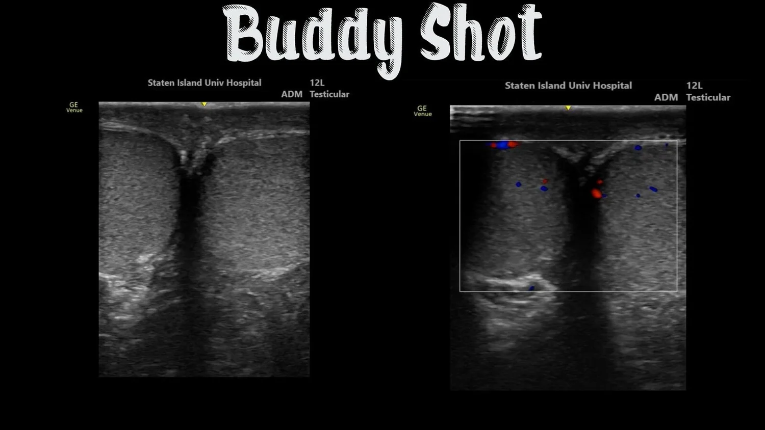
1. The buddy shot
Place both testes side by side in a grayscale image and a corresponding color Doppler image, and compare the two sides.
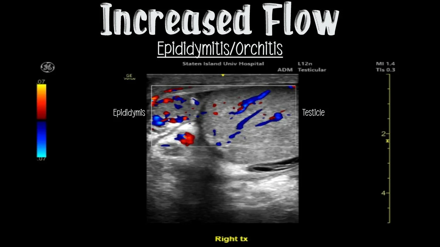
2. Increased flow: epididymitis and orchitis
Color Doppler shows increased flow in epididymitis/orchitis. The epididymis and testicle are labeled separately in the same image.
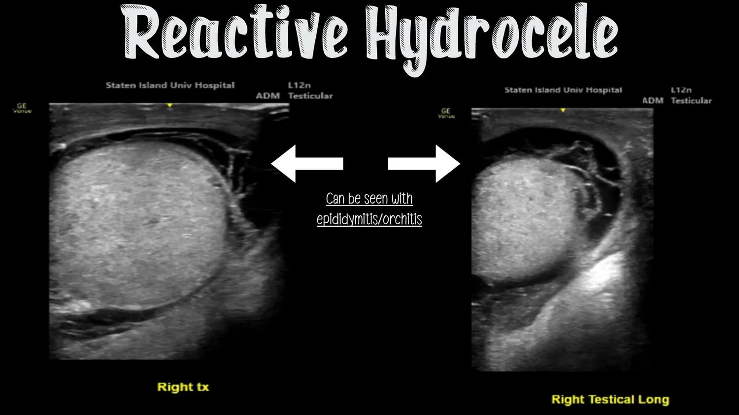
3. Reactive hydrocele
Two grayscale views, with arrows highlighting fluid around the testicle. A reactive hydrocele can be seen with epididymitis/orchitis.
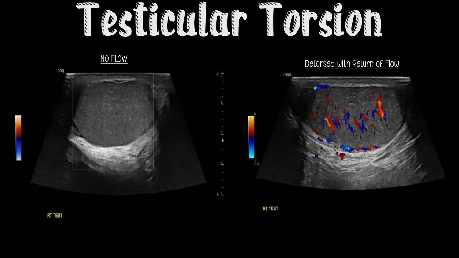
4. Testicular torsion: before and after detorsion
A torsed testicle with no flow on color Doppler, and the same testicle after detorsion with return of flow.
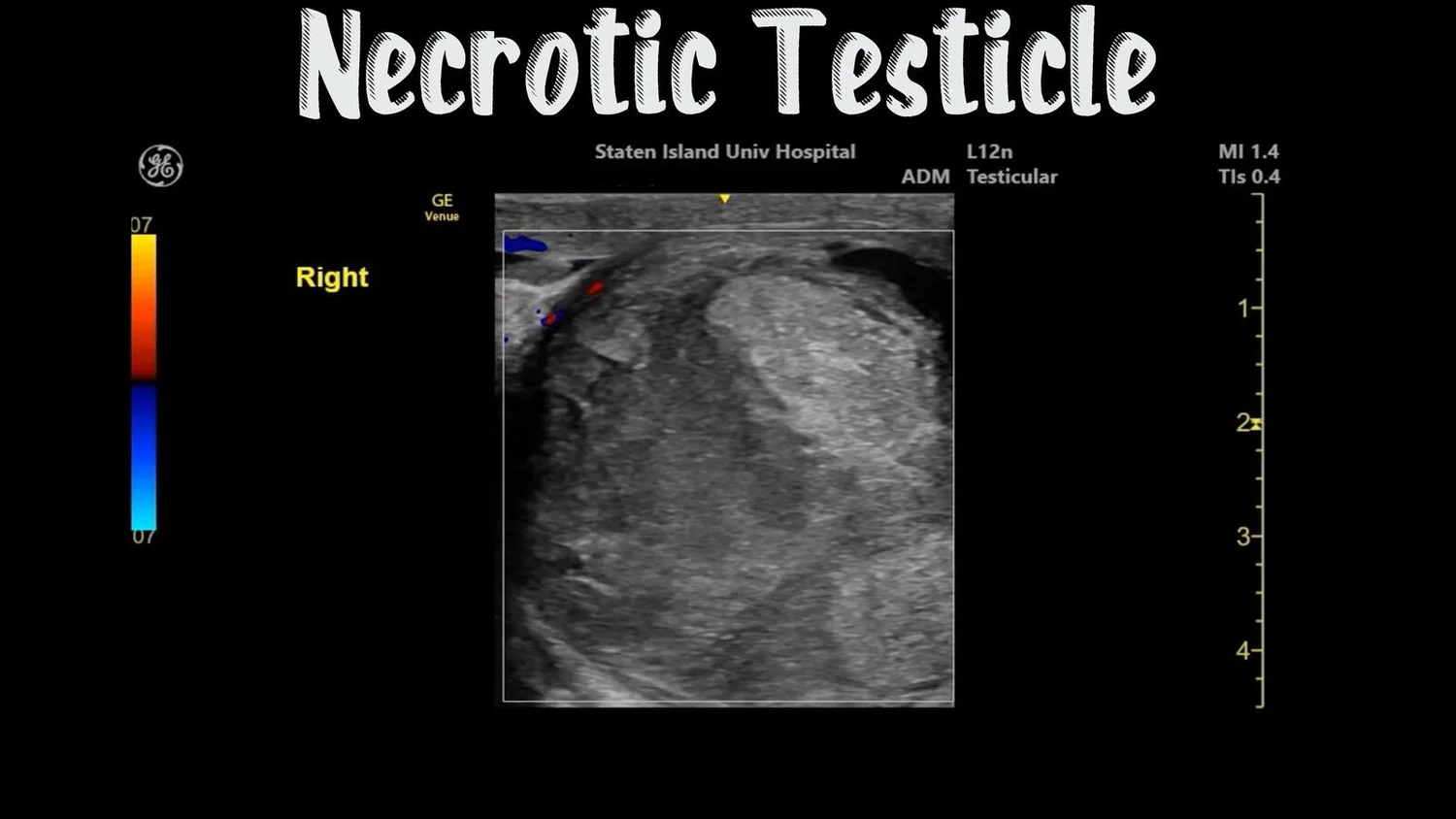
5. Necrotic testicle
Ultrasound appearance of a necrotic testicle.
Practical teaching points
Review the paired grayscale and color Doppler buddy-shot views.
Identify the separately labeled epididymis and testicle in the increased-flow example.
A reactive hydrocele can be seen with epididymitis/orchitis.
Compare the no-flow torsion image with the return-of-flow image after detorsion.
Review the final necrotic-testicle example alongside the preceding images.
Resource
Beyond the Twist: Ultrasound differentiation of epididymitis, orchitis and torsion (PDF, 6 pages)
Ultrasound image gallery