

9/30/25:
Ultrasound Education for the Week
Dr. Lea Salazar, MD
Chief Complaint:
Facial Swelling
HPI:
45 yo M with pmh of HTN, HLD and GERD, presents with left-sided facial swelling x 2 days. Patient says pain is worse at the upper jaw and radiates to his left ear. Patient denies dental pain, but admits pain is worse with eating. No difficulty swallowing or pain on swallowing. No voice changes. No difficulty breathing. Denies fever or chills.
Physical Exam:
Focused exam: L sided facial swelling, mildly indurated, no skin changes. Swelling extends from area anterior to the L ear to the angle of the mandible. Mildly ttp. Dentition normal. No gingival swelling. OP open and patent, no tonsillar swelling or erythema. No tongue or sublingual swelling. Bilateral external auditory canals normal, bilateral TMs normal.
Longitudinal
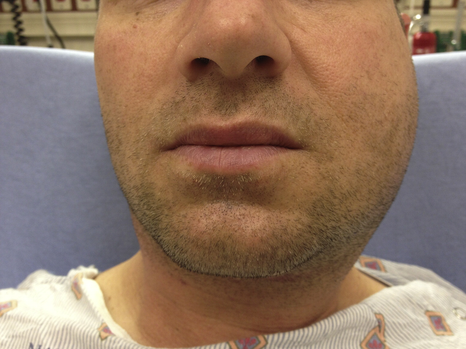
What’s your differential?
You grab the ultrasound. What organ will you be evaluating?
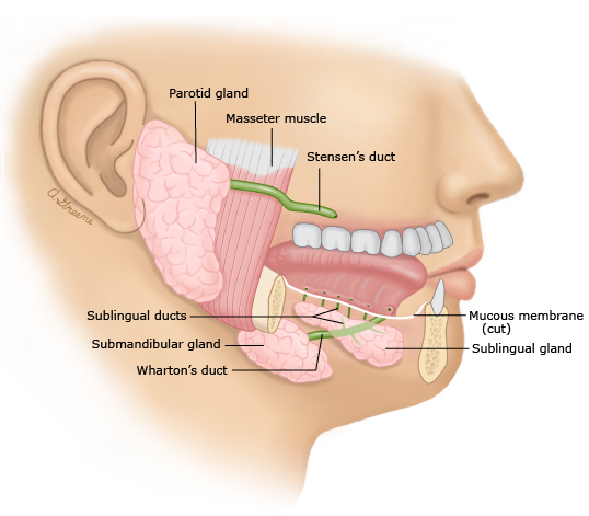
Based on the history and the location of the swelling, let’s examine the parotid gland
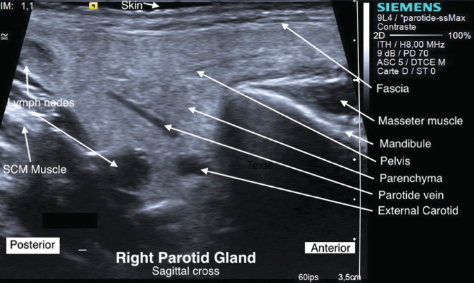
Anatomy
POCUS technique:
Which probe do you use?
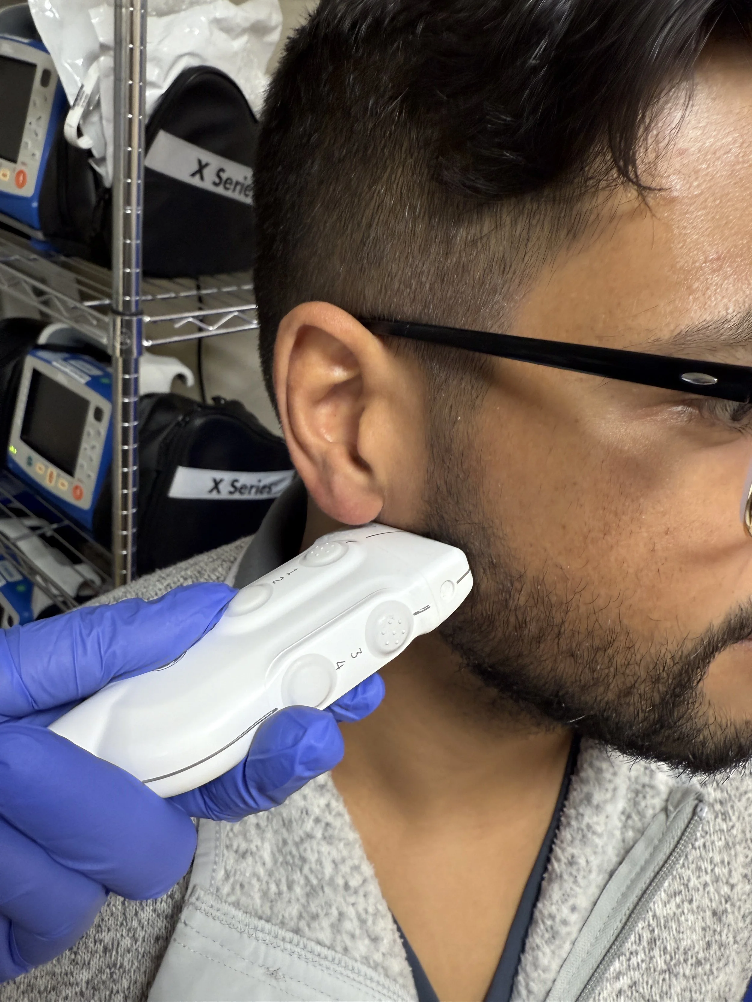
The high-frequency, linear probe.
Where do you place the probe?
Anterior to the ear and superior to the angle of the mandible
Scan in 2 views, the longitudinal and the transverse (cross) views:
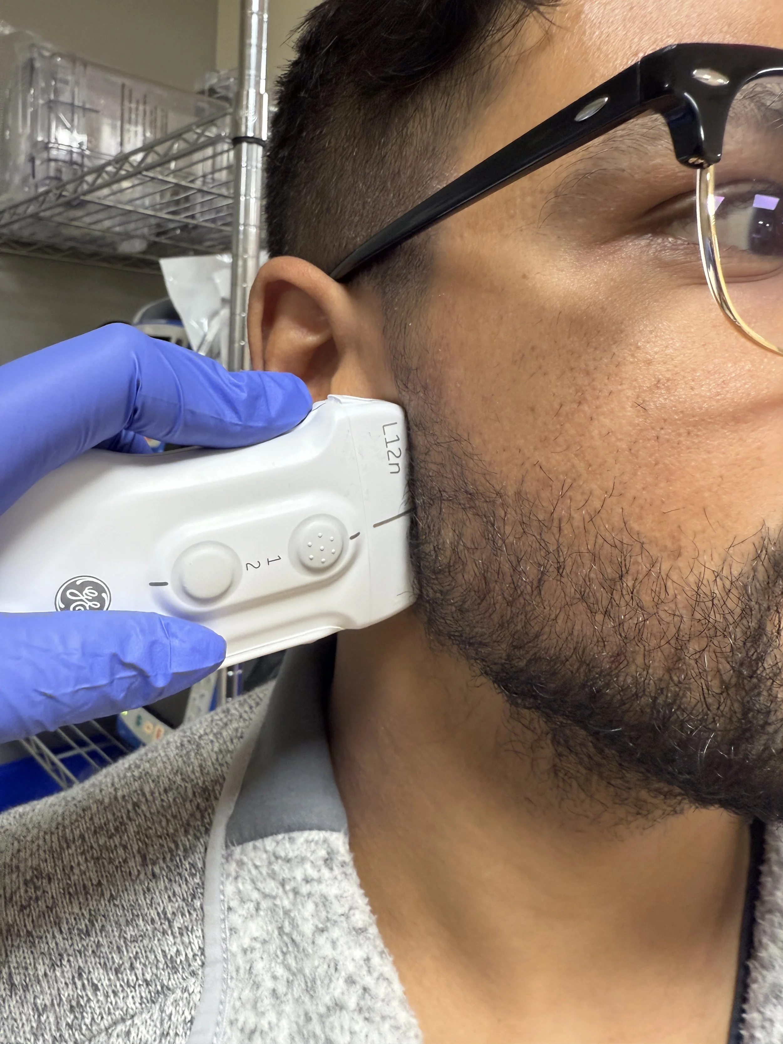
Longitudinal
Remember to scan both parotid glands for comparison.
Normal parotid ultrasound:
1. Hyperechoic organ with homogeneous parenchyma
2. Usually cannot easily visualize parotid duct (Stensen’s duct)
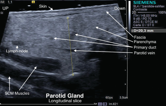
Transverse
Now back to our patient…
Transverse
Longitudinal
Transverse
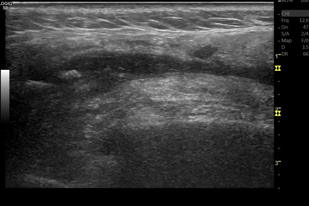
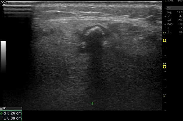
What does your scan show?
Dilated parotid duct
Shadowing stone
What is the patient’s diagnosis?
Parotid sialolithiasis
What is the management?
Hydration
Warm compress
Massaging the gland and “milking” the duct
Foods that promote salivary flow (citric food, lemon drops, sour candies, etc)
If concerned about secondary infection, start Augmentin.
Resources:
https://radiopaedia.org/cases/parotid-duct-stone-3
Salivary Gland Diseases Ultrasound by Phillippe Katz