

8/26/25:
Ultrasound of Central Retinal Artery Occlusion
Dr. Josh Greenstein, MD
Case:
A 71-year-old female with a history of hypertension and atrial fibrillation presented to the ED at 12:08 pm with sudden painless loss of vision in her left eye for 45 minutes. On exam, visual acuity was light perception only in the affected eye. A bedside ocular ultrasound was performed (see below), and fundoscopic exam was not attempted.
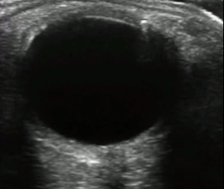
Normal Right Eye
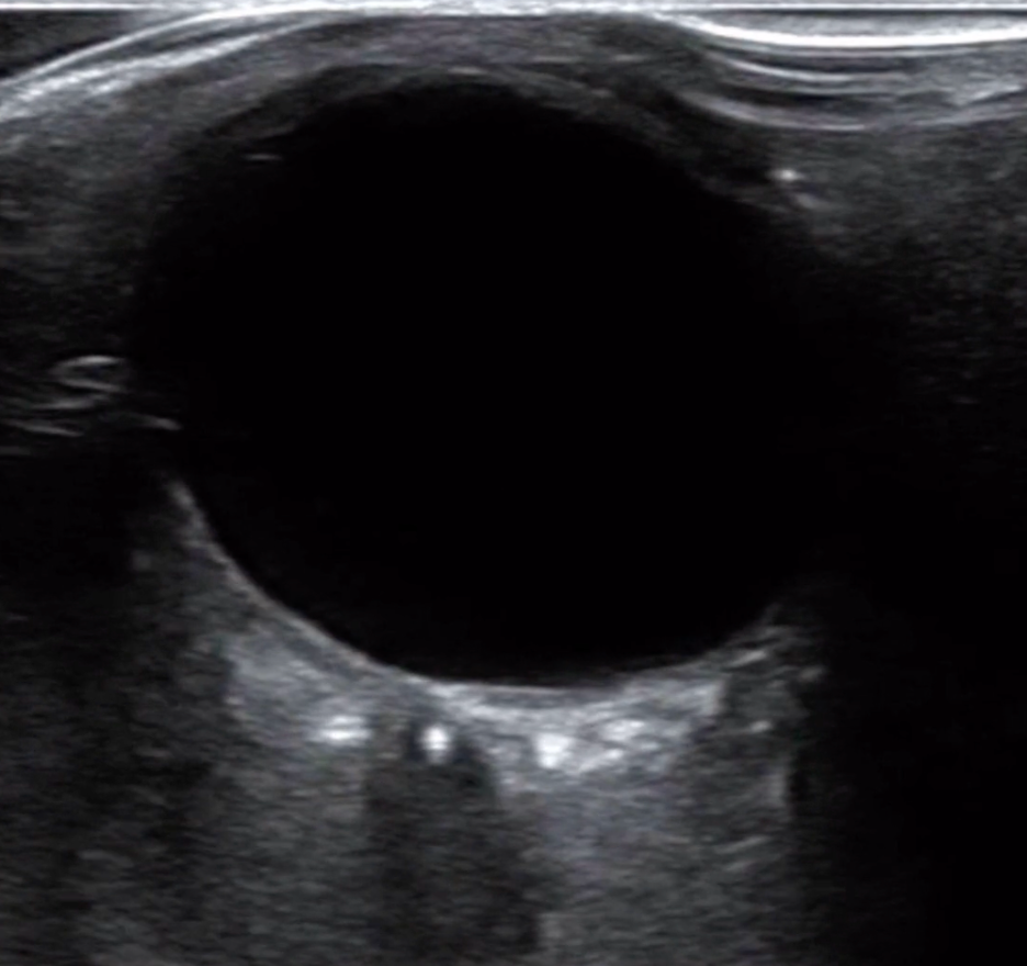
Abnormal Left Eye
What is the diagnosis?
Central Retinal Artery Occlusion (CRAO)
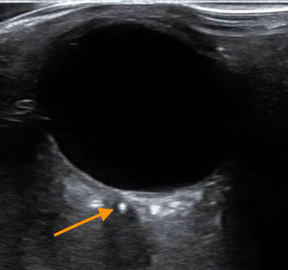
The retrobulbar spot sign (RBSS) is an ocular ultrasound finding seen in cases of CRAO. It appears as a hyperechoic focus within the lumen of the central retinal artery, located posterior to the globe, and represents an intraluminal embolus obstructing blood flow. This sign is considered highly specific for thromboembolic CRAO, as it directly visualizes the embolic material causing the occlusion.
Detection of the retrobulbar spot sign can support rapid diagnosis when fundoscopic examination is inconclusive.
Can the RBSS indicate any other pathology?
The RBSS retrobulbar spot sign is most classically associated with an embolic CRAO, but it is not entirely specific. Other conditions that may produce a similar finding include:
Optic Nerve Drusen - tiny deposits of calcium, protein that accumulate within the optic nerve
Artifact – may mimic a RBSS
Why is this important?
The RBSS has high specificity but low sensitivity for CRAO. In practice, this means:
If the RBSS is seen on ultrasound and the patient’s clinical picture matches sudden painless monocular vision loss, cherry-red spot on fundoscopy (if seen), the diagnosis of embolic CRAO is very likely.
Low sensitivity → Many patients with CRAO will not show this sign on ultrasound, so its absence does not rule out the condition.
How do you get a CRAO?
CRAO is most commonly caused by an embolus from carotid artery atherosclerosis or cardiac sources (e.g., atrial fibrillation, valvular disease). Less common causes include giant cell arteritis, hypercoagulable states, or trauma-induced arterial occlusion.
What are some risk factors for CRAO?
Hypertension
Diabetes Mellitus
Carotid artery stenosis
Atrial fibrillation
Smoking
Giant cell arteritis
What are the symptoms?
Sudden, painless, monocular vision loss
Relative afferent pupillary defect (RAPD)
Sometimes patients report transient visual loss (amaurosis fugax) prior to the occlusion
What is the differential diagnosis?
Branch retinal artery occlusion
Central retinal vein occlusion
Retinal detachment
Vitreous hemorrhage
Ocular ischemic syndrome
Optic neuritis (is painful)
When concerned for CRAO, do I need imaging?
Funduscopic exam is usually diagnostic. Ocular ultrasound may be used adjunctively.
What is the treatment?
CRAO is an ophthalmic emergency. Immediate measures include:
Emergent ophthalmology consultation
Ocular massage (to attempt dislodging embolus)
Consideration of TNK in discussion with neurology and ophtho
Consideration of IR for embolectomy.
Evaluate for underlying embolic source and initiate secondary prevention