

Ultrasound of the neck: a painful anterior neck mass
History
An 11-year-old previously healthy girl, up to date on vaccines, presents with her mother for a painful anterior neck mass. She has had a tiny bump on the front of her neck for almost a year, but over the past few weeks it has grown significantly and become painful. There is no fever, nausea or vomiting, or respiratory distress. She has mild discomfort with swallowing.
This case is drawn from the author's own experience as an intern.
Exam
Speaking in full sentences and tolerating secretions.
Normal oropharynx.
Bilateral breath sounds without stridor or wheezing.
Anterior neck mass.
Ultrasound
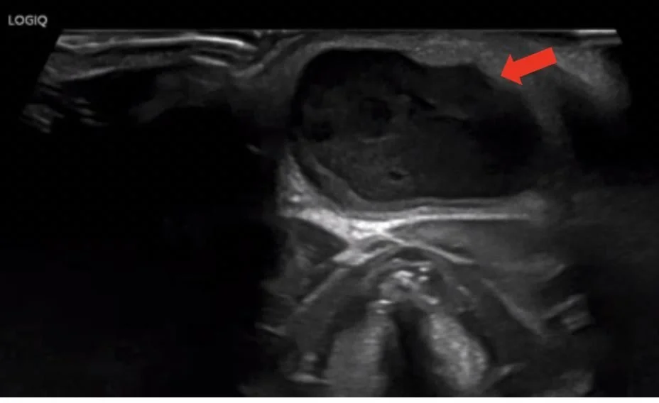
A paramedian, complex cystic structure at the level of the thyroid cartilage, with surrounding inflammatory change.
Diagnosis and disposition
Infected thyroglossal duct cyst. She was started on IV antibiotics and admitted.
The case itself is straightforward, but it is a good opportunity to review ultrasound of the neck.
Scanning technique
Probe: a high-frequency linear probe (low penetration, high resolution).
Position: patient supine with the neck extended. Scan in both longitudinal and transverse planes.
Normal anatomy
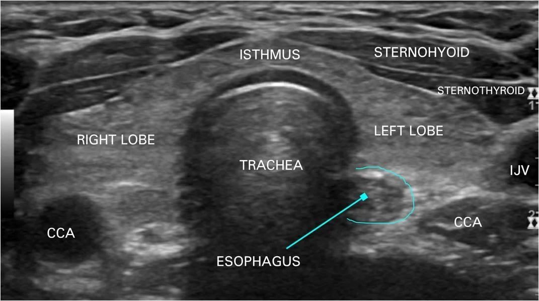
Thyroid: bilateral lobes joined across the trachea, with a homogeneous ground-glass appearance, hyperechoic relative to the adjacent strap muscles.
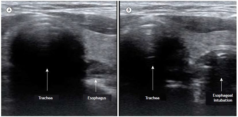
Trachea: a bright hyperechoic line (an upside-down U), with distal shadowing from the air–mucosa interface.
Esophagus: a collapsed round or oval structure, posterolateral and to the patient's left of the trachea. Normally it has no air or shadowing.
Congenital neck cysts
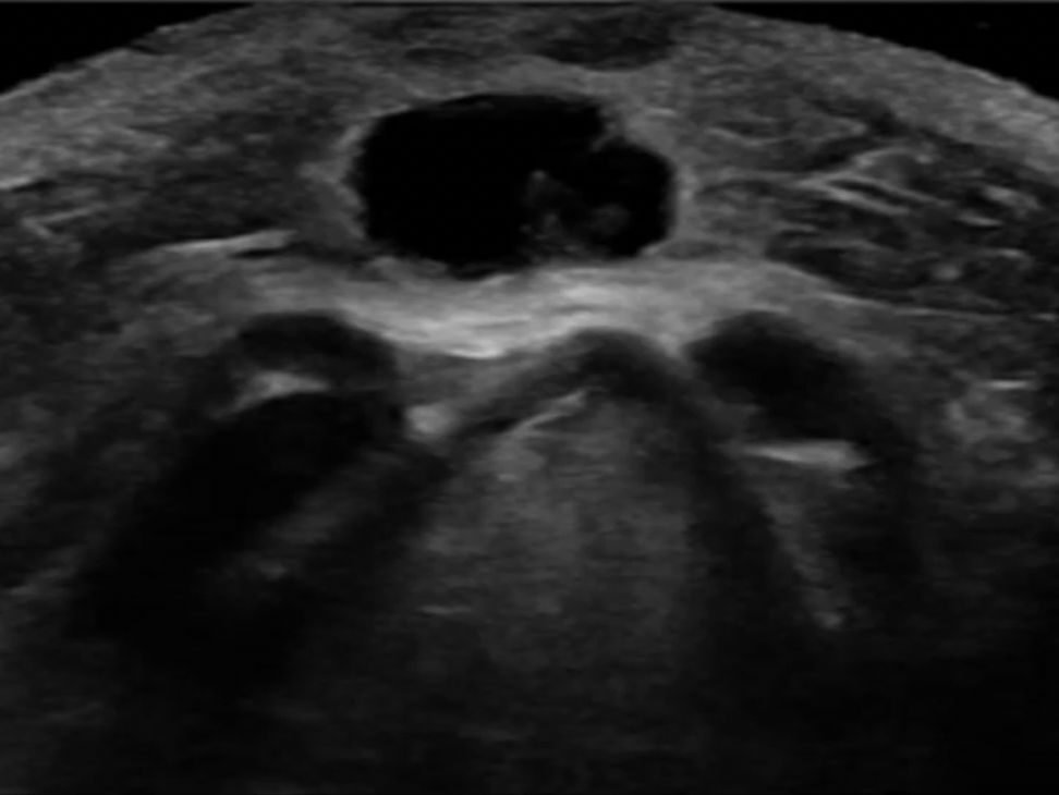
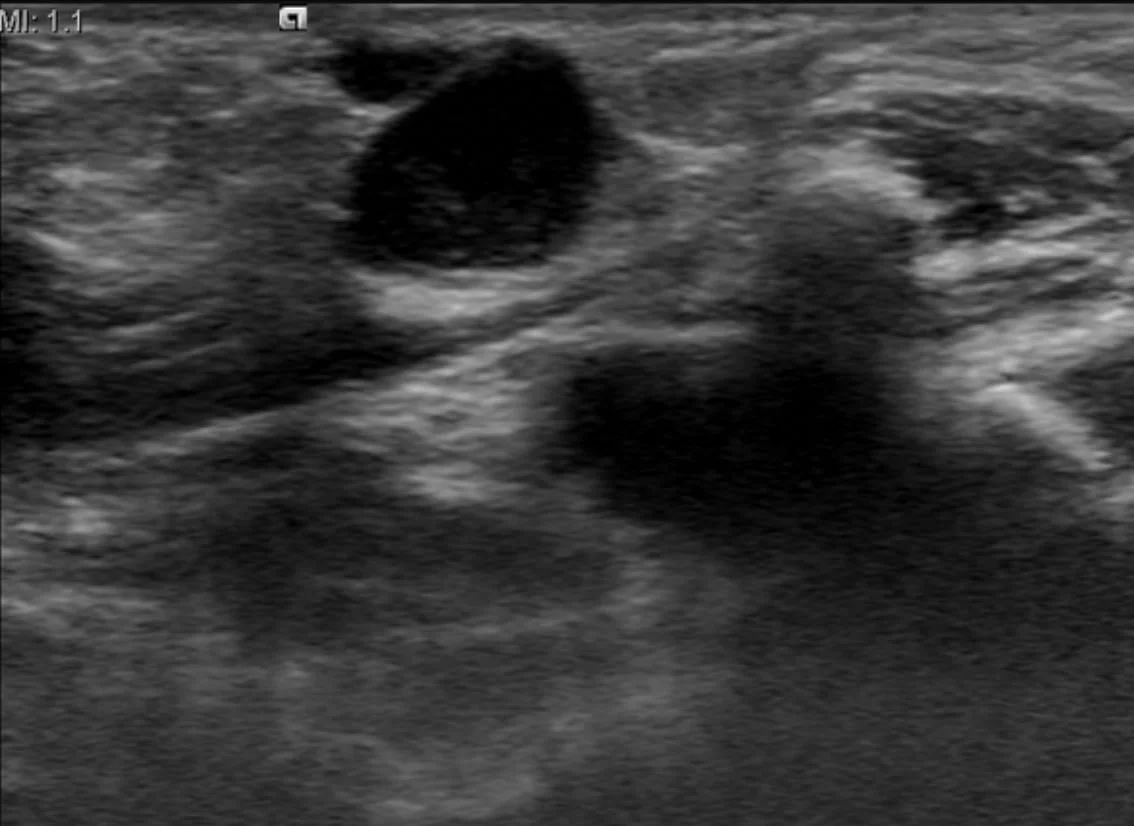
| Thyroglossal duct cyst | Branchial cleft cyst | |
|---|---|---|
| Location | Usually midline, most at or below the hyoid | Lateral neck, usually anterior to the sternocleidomastoid |
| Appearance | Well-circumscribed, anechoic, fluid-filled, no internal vascularity | Well-circumscribed, anechoic, fluid-filled, no internal vascularity |
Infection can make either cyst appear complex, with surrounding inflammatory change, as in this case.
Bonus applications
Endotracheal tube confirmation: "Did you tube the goose?"
Tube in the trachea: a single air–mucosa interface (the trachea), with increased posterior shadowing.
Tube in the esophagus: a "double trachea" (double tract) sign.
Thyroid nodules
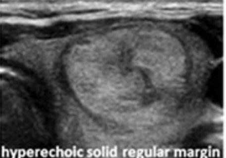
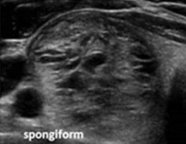
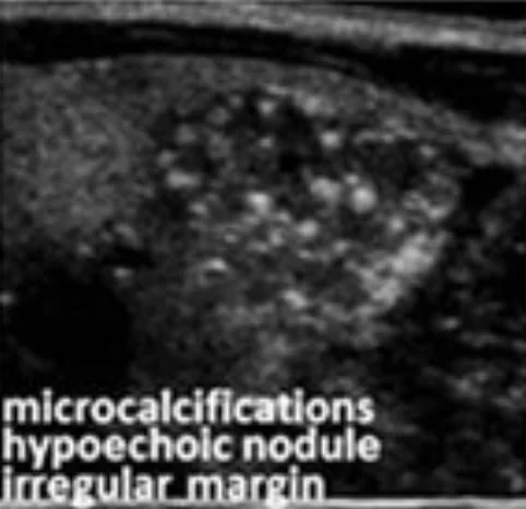
Appearances vary.
Higher suspicion for malignancy: microcalcifications, irregular borders, or high internal blood flow.
Refer for outpatient comprehensive thyroid ultrasound.
Clinical pearls
Midline means thyroglossal; lateral means branchial.
Use the linear probe, extend the neck, and scan in two planes.
A double-tract sign on neck ultrasound after intubation means an esophageal intubation.