

The popliteal sciatic nerve block ("pop block")
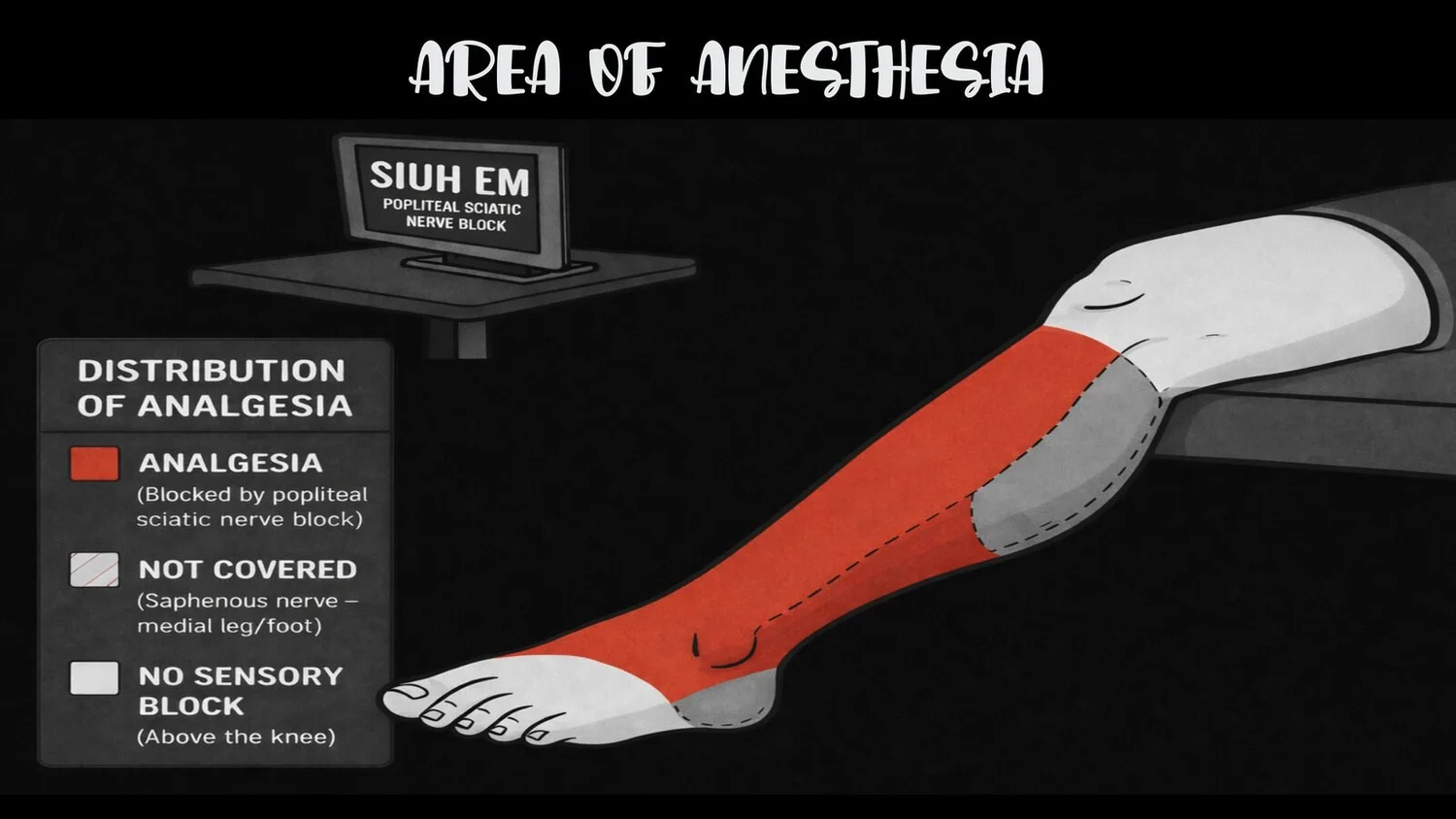
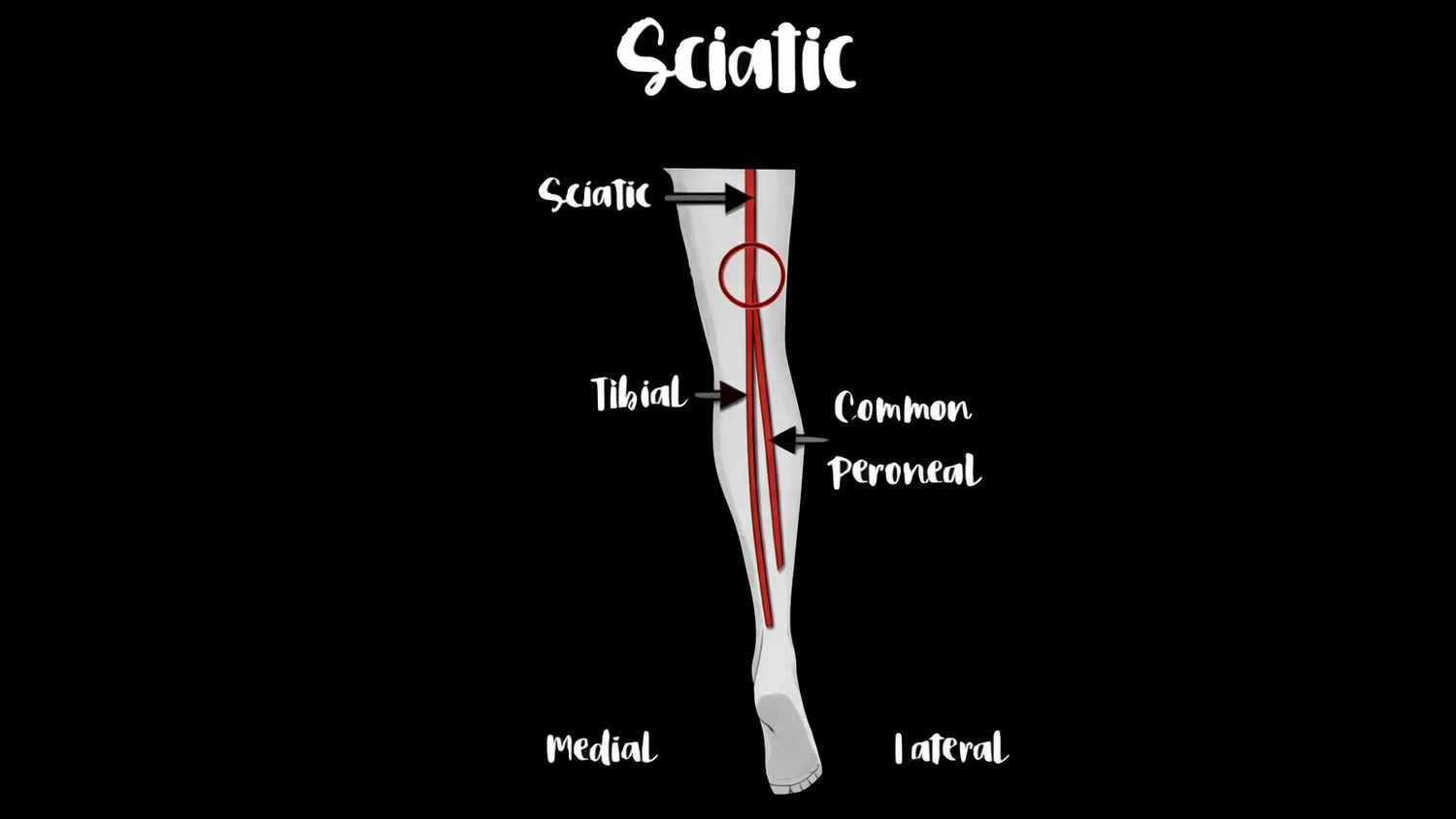
An ultrasound-guided block of the distal sciatic nerve in the popliteal fossa, near where it divides into the tibial and common fibular (peroneal) nerves. It provides analgesia to much of the lower leg and foot.
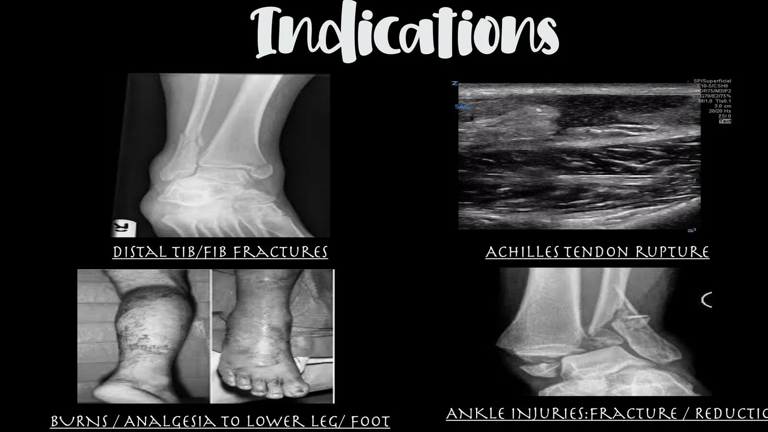
Indications
Distal tibia/fibula fractures
Achilles tendon rupture
Ankle injuries: fracture analgesia and reductions
Burns, and analgesia of the lower leg and foot
Area of anesthesia and anatomy
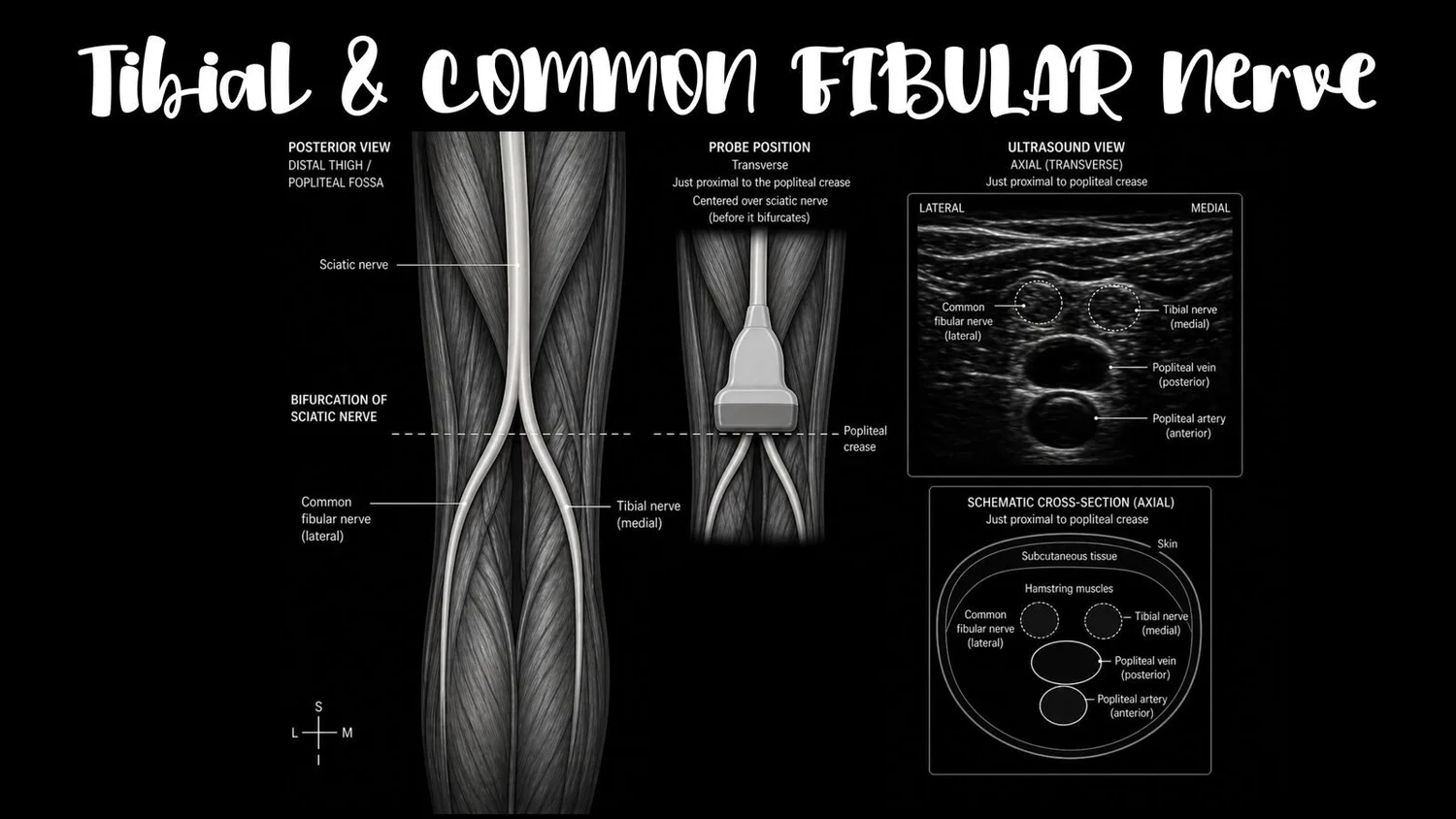
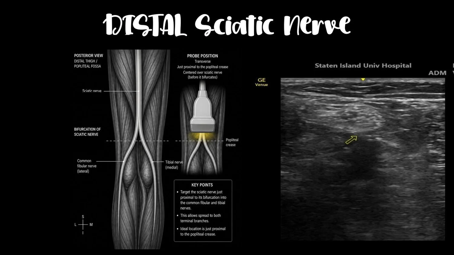
Target: the distal sciatic nerve, injected around the nerve at or just above its bifurcation into the tibial and common fibular nerves.
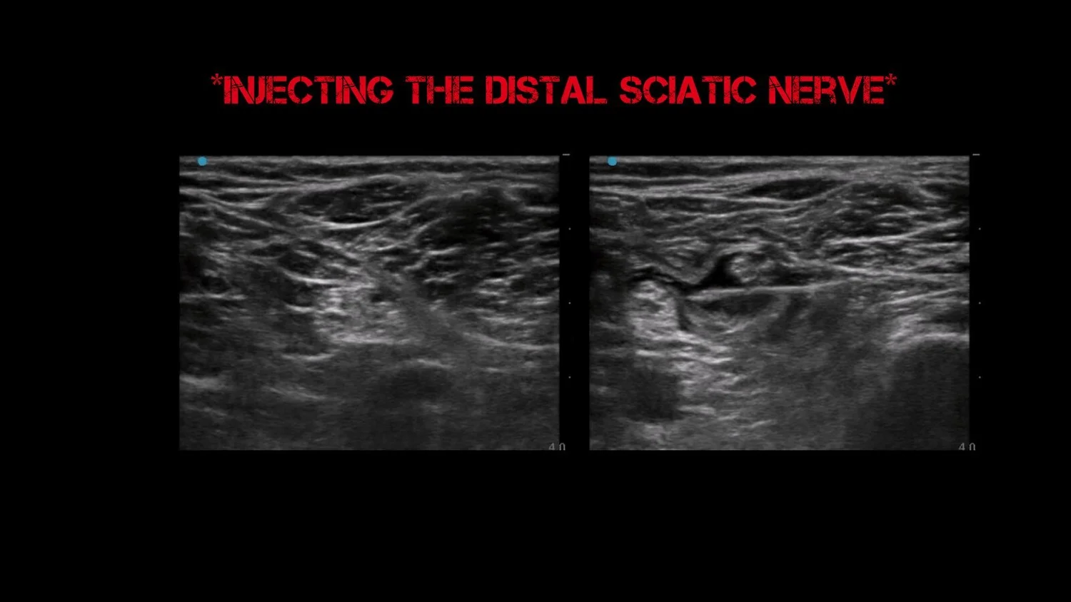
The figures below illustrate the area of anesthesia, the anatomy, and the sonographic target for injection.
Technique
Position: prone or lateral (posterior approach), or supine with the leg elevated (lateral approach).
Probe: linear transducer, transverse in the popliteal crease.
Find the anatomy: popliteal artery (about 3–4 cm deep) with the vein just superficial to it; the tibial nerve sits superficial and lateral to the vein, and the common peroneal nerve is more superficial and lateral still. Scan proximally to where the two join (usually 5–10 cm above the crease).
Needle: in-plane, aiming for the common sheath around both nerves at the level where they begin to separate.
Inject: 15–20 mL of local anesthetic. Give 1–2 mL first to confirm spread around both nerves; stop if you feel high resistance.
Clinical pearls
A single block can spare the patient opioids during ankle reductions and painful lower-leg injuries.
Assess compartment syndrome risk before you block. In high-risk injuries such as tibial shaft fractures, coordinate with orthopedics, because a dense block can mask the pain of evolving compartment syndrome.
Document a full neurovascular exam before the block.