

POCUS on the brain: cracking the case of pediatric skull fractures
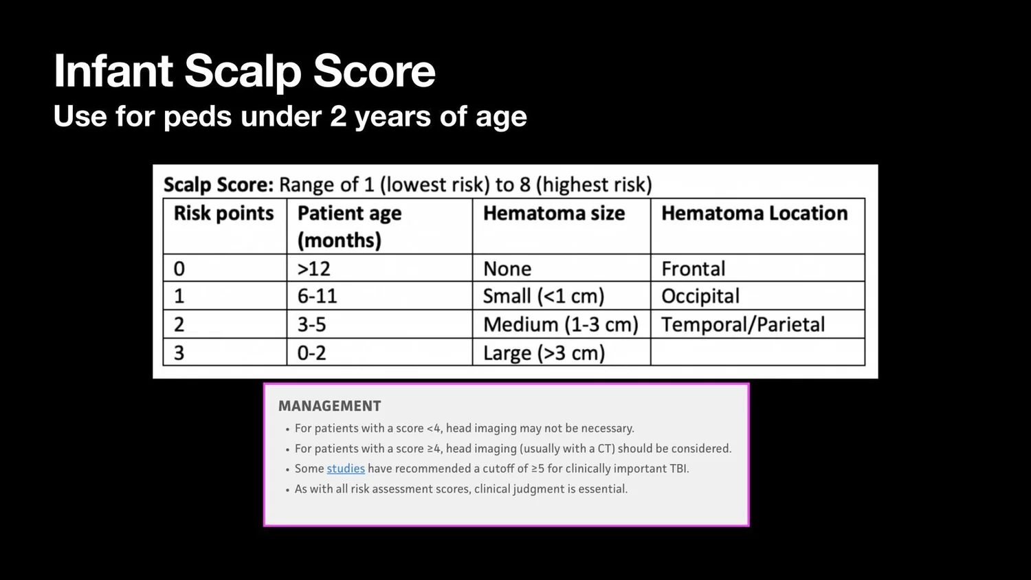
Consider point-of-care ultrasound (POCUS) when you evaluate scalp hematomas in children with head injury. For children under 2 years, review the Infant Scalp Score (shown below) to help decide whether further imaging is warranted.
When to use skull ultrasound
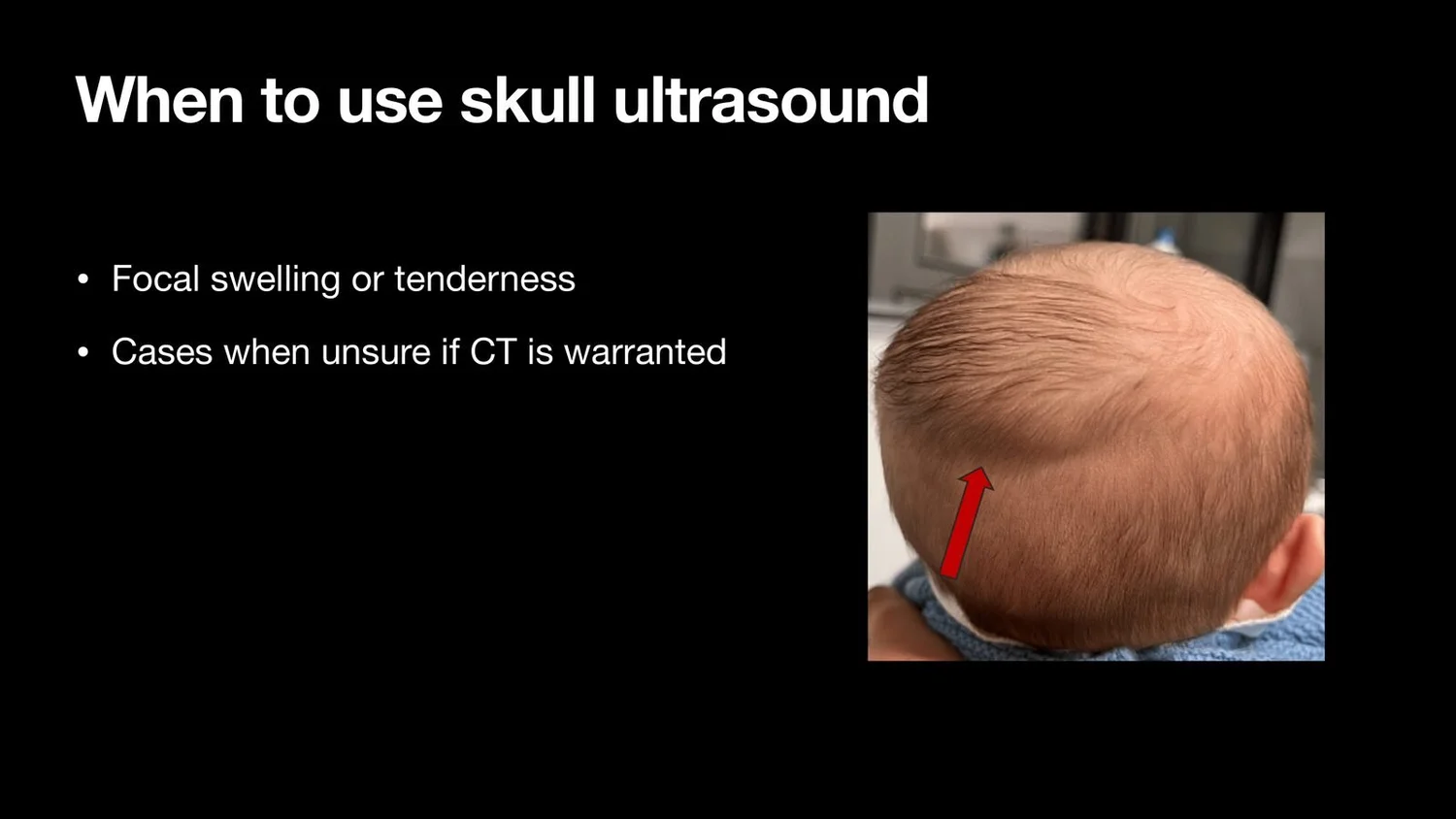
Focal swelling or tenderness: scalp hematomas or focal swelling, and palpable step-offs or tenderness.
A mechanism concerning for skull injury.
When it is unclear whether CT is warranted, for example in intermediate-risk patients.
Technique ("so easy, even for the novice")
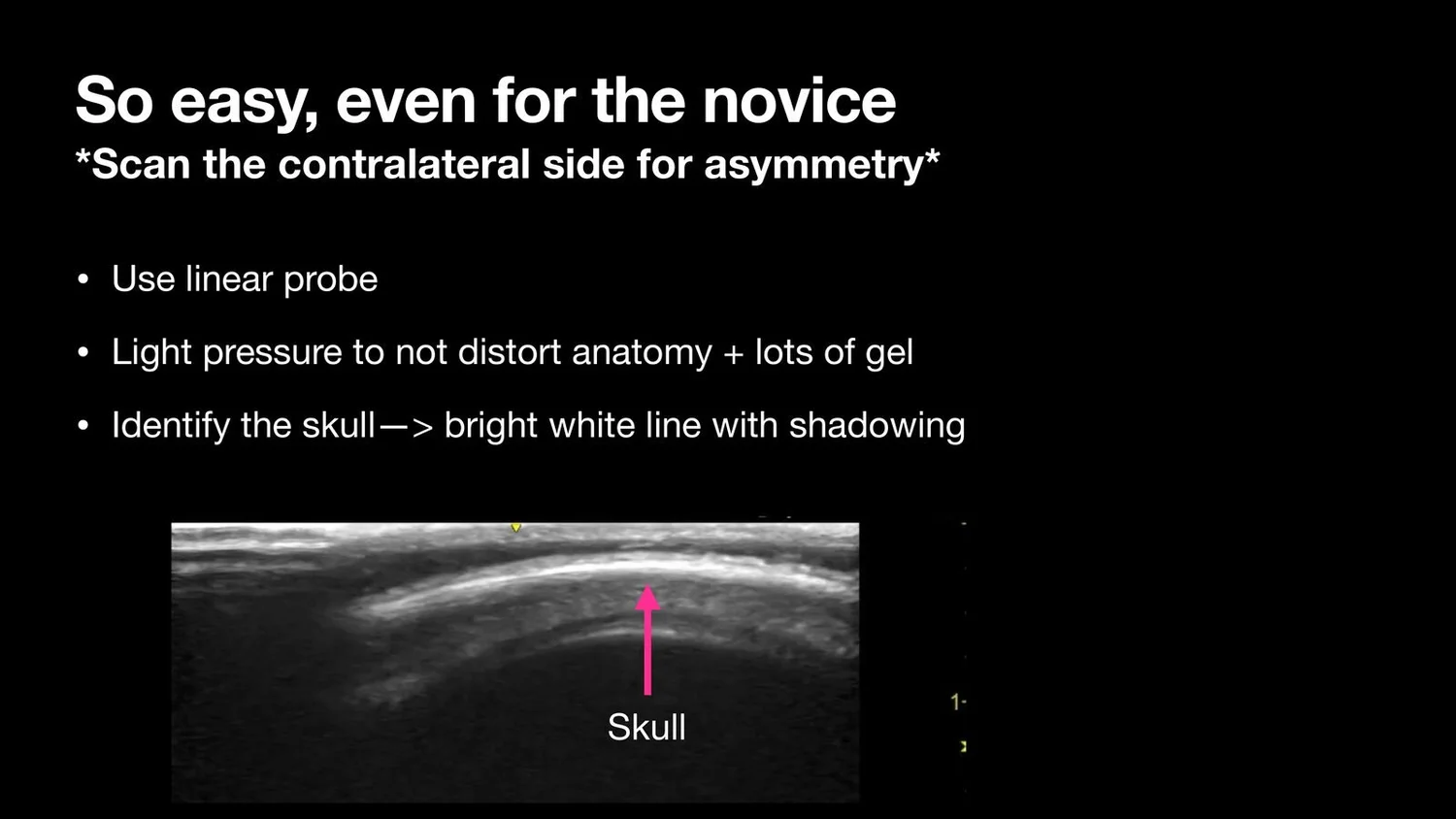
Use the linear probe.
Apply light pressure and plenty of gel so you do not distort the anatomy.
Identify the skull: a bright white (hyperechoic) line with posterior shadowing.
Scan the contralateral side to compare for asymmetry.
Fracture or suture?
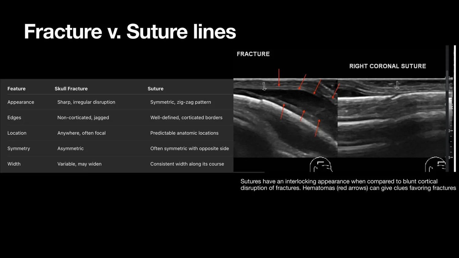
Sutures have an interlocking, symmetric appearance.
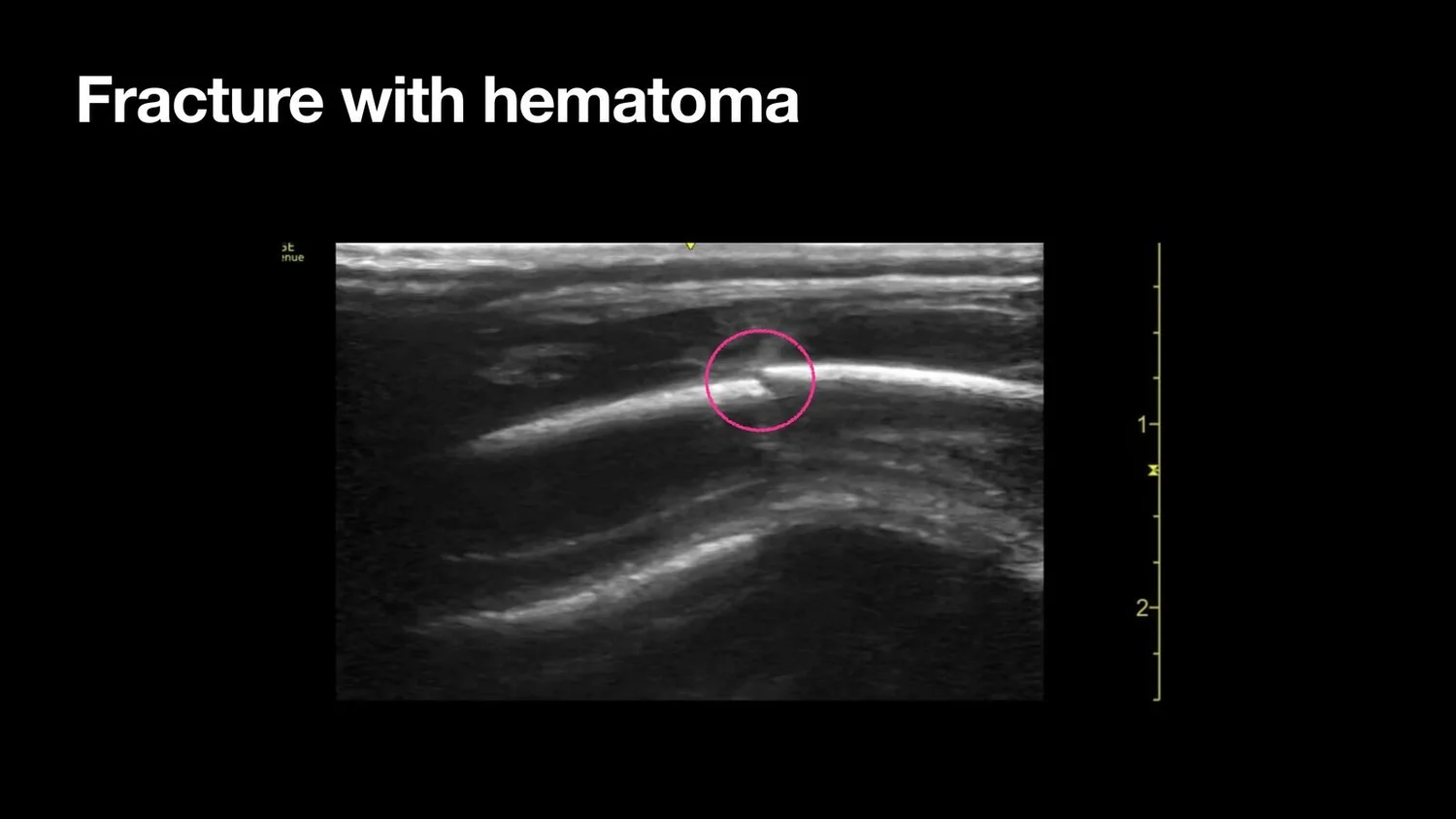
Fractures show a blunt disruption of the cortex.
An overlying hematoma is a clue that favors fracture.
How POCUS helps
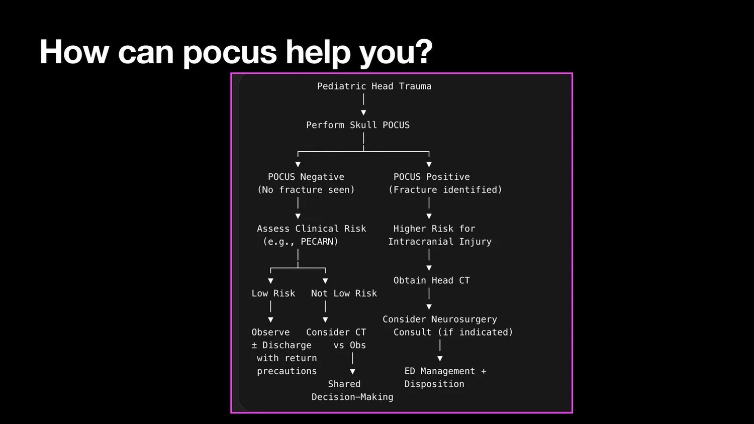
POCUS gives a rapid, radiation-free look at the skull under a hematoma. Combined with the clinical assessment, a validated decision rule, and the Infant Scalp Score for children under 2, it can help you decide which children need CT.
Clinical pearls
Always compare with the other side. Sutures are paired and symmetric; fractures are not.
Do not compress the hematoma. Light pressure and lots of gel give the best image.
A fracture on POCUS changes your risk assessment. Combine it with the clinical picture to decide on further imaging.