

Pediatric ultrasound: hypertrophic pyloric stenosis
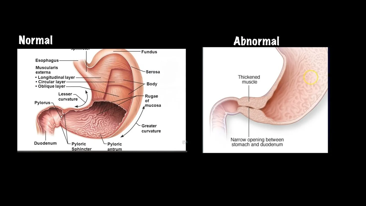
This pediatric ultrasound edition covers hypertrophic pyloric stenosis (HPS). Use the guide (figures below and linked PDF) to perform the exam yourself or interpret the technician's images. You may avoid signing the patient out to the next team while waiting for a preliminary read.
When to consider pyloric stenosis
Age: 2 weeks to 2 months
History and exam:
Non-bilious emesis after feeds
Infant still hungry after vomiting
Weight loss or failure to thrive
Scanning technique
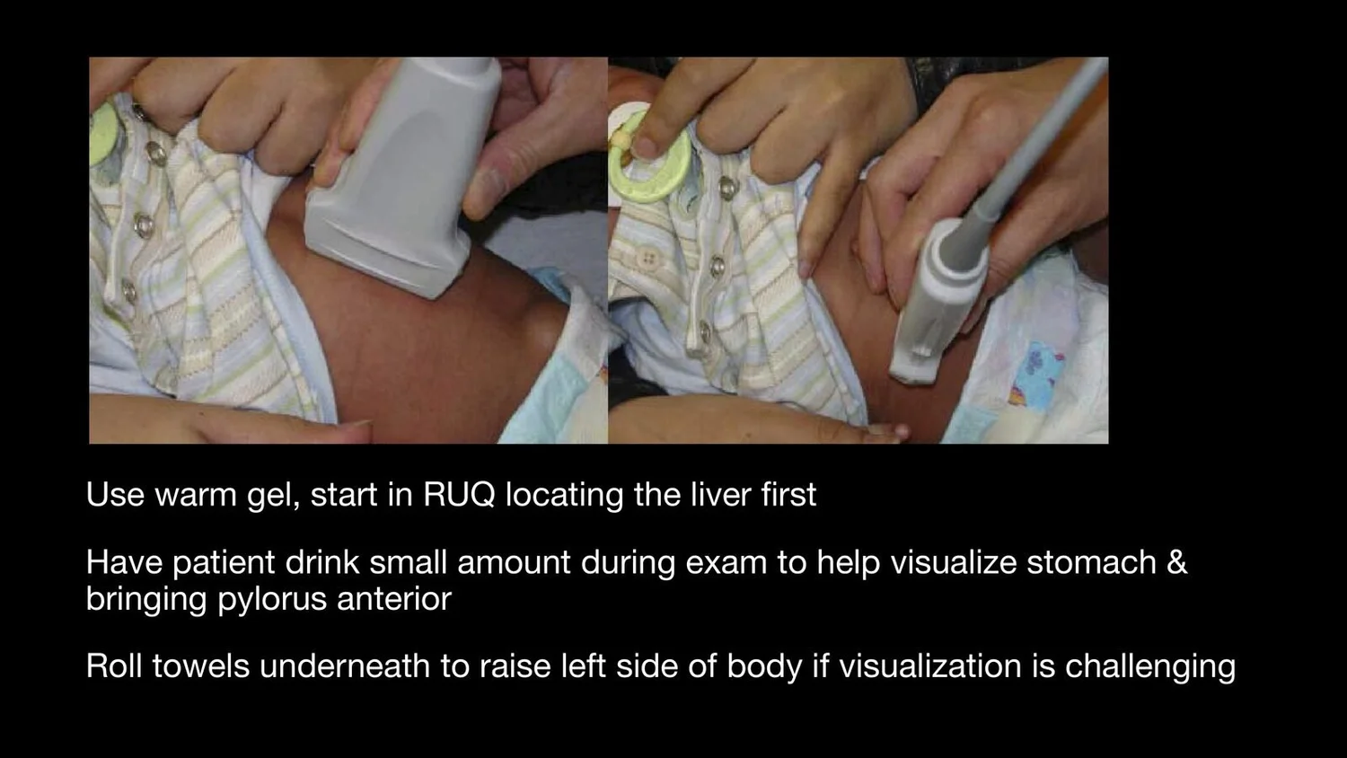
Use warm gel.
Start in the right upper quadrant and find the liver first, then follow toward the gallbladder and pylorus.
Have the infant drink a small amount during the exam. This helps visualize the stomach and brings the pylorus anteriorly.
If visualization is difficult, roll towels under the left side of the body to raise it.
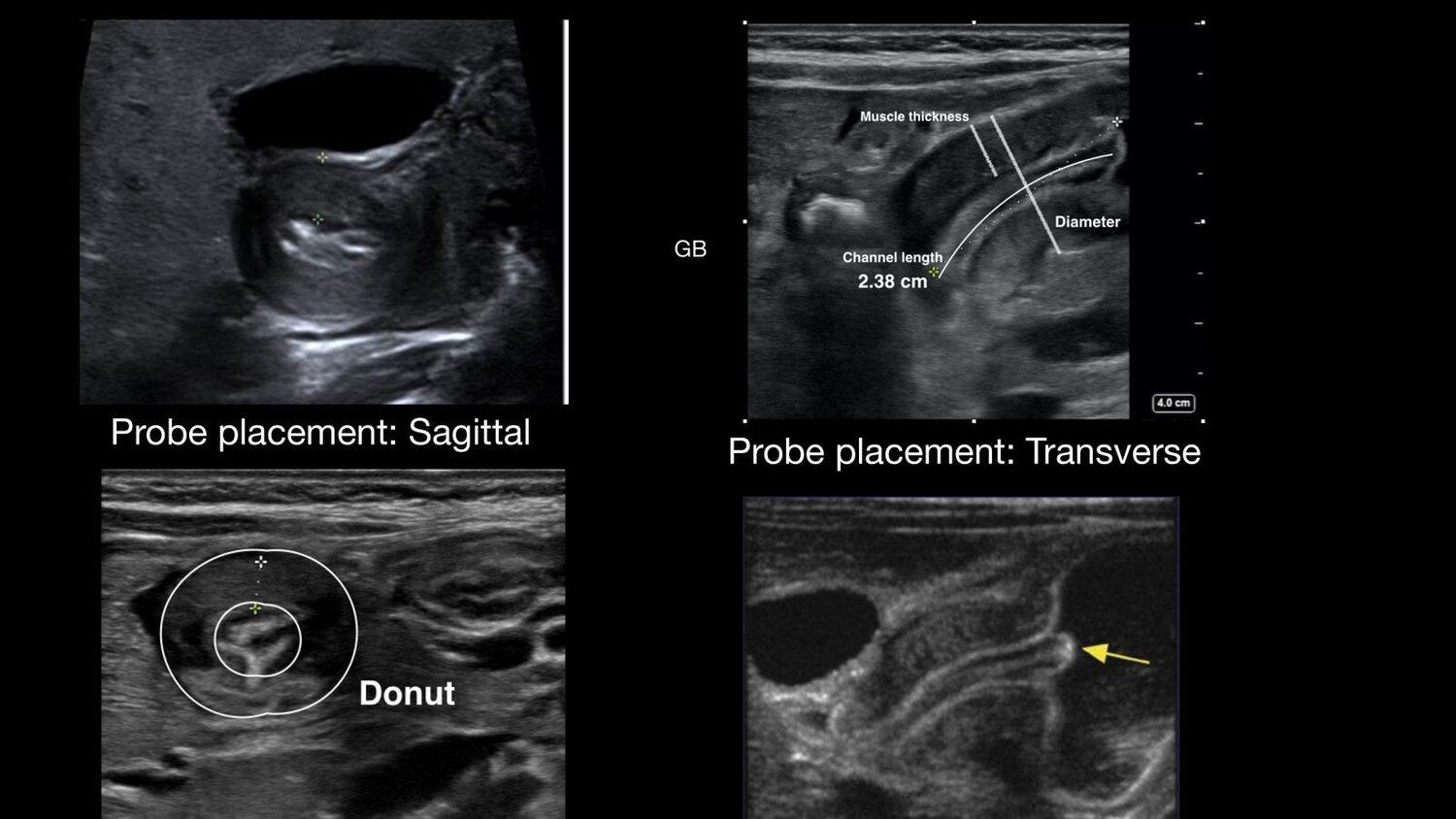
Image the pylorus in both sagittal (long-axis) and transverse probe orientations. Compare normal and abnormal appearances. A dilated stomach supports obstruction.
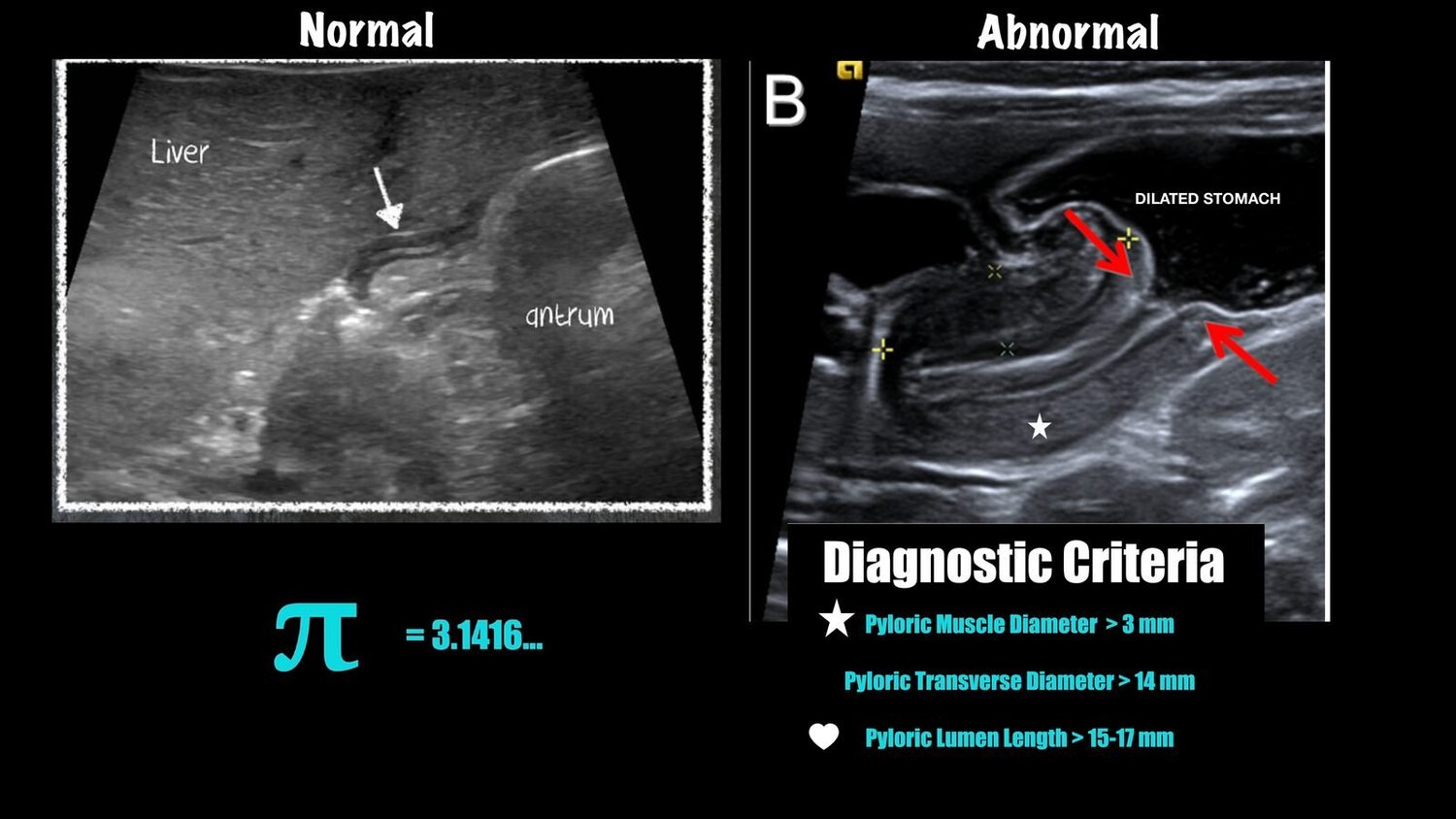
Abnormal (pyloric stenosis) measurements: pyloric muscle thickness > 3 mm; pyloric transverse diameter > 14 mm; pyloric channel length > 15–17 mm.
Tama's tips
Beware pylorospasm. Temporary muscle thickening can mimic HPS. Watch over time.
Look for gastric contents passing through the pyloric channel. Fluid passing freely argues against fixed obstruction.
Check electrolytes. HPS classically causes hypochloremic, hypokalemic metabolic alkalosis.