

When time is money: identifying air on ultrasound
Gas in soft tissue or in the gallbladder wall almost always means a surgical emergency. Bedside ultrasound can show it within minutes, well before a formal study is done and read. This edition reviews two ED cases where point-of-care ultrasound (POCUS) made the diagnosis. You do not need to be an ultrasound expert to recognize these findings.
Case 1: Necrotizing fasciitis (Fournier's gangrene)
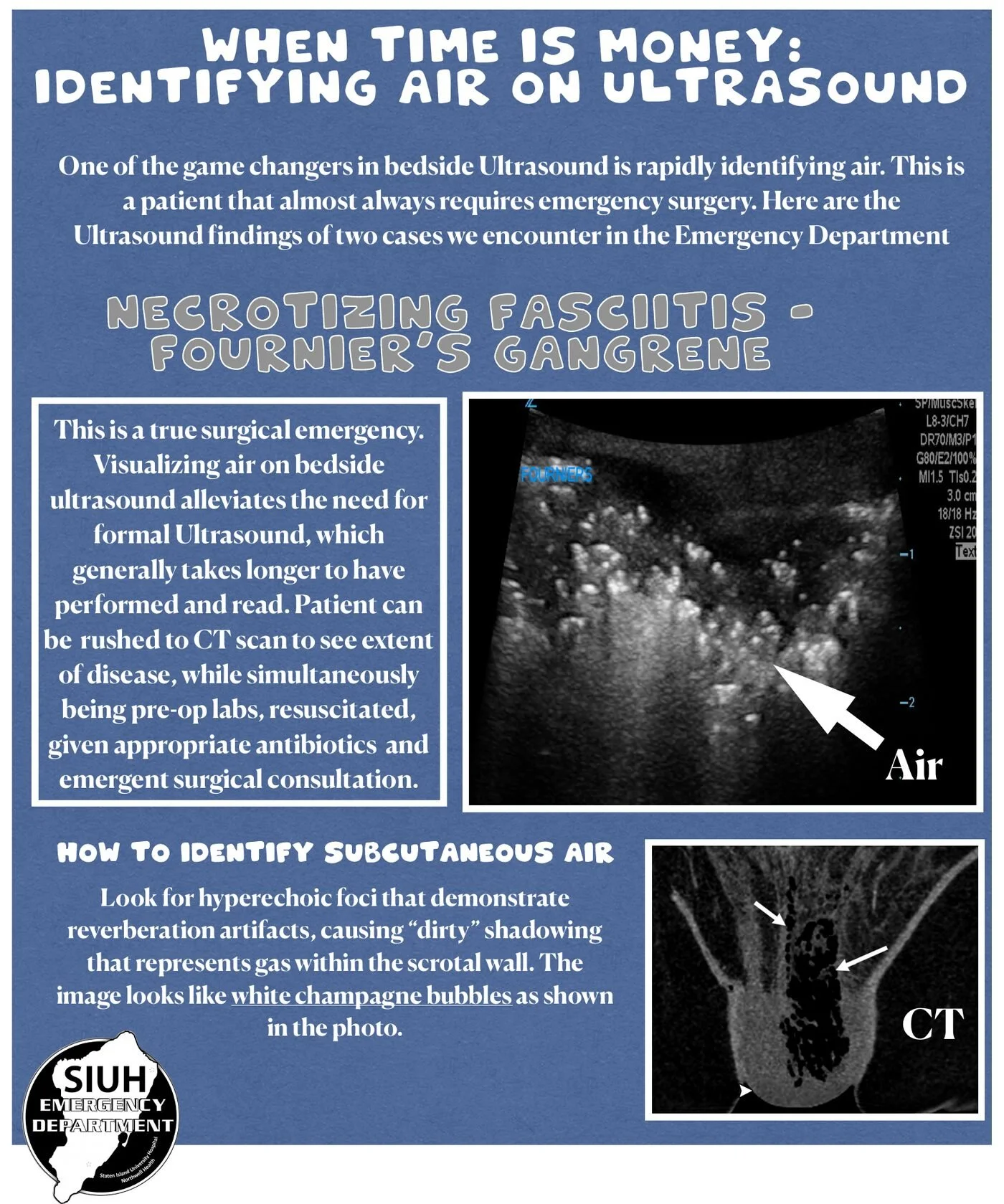
Fournier's gangrene is a true surgical emergency. Seeing gas in the scrotal or perineal soft tissues on bedside ultrasound removes the need to wait for a formal ultrasound, which generally takes longer to be performed and read.
How to identify subcutaneous air
Look for hyperechoic foci with reverberation artifact that cause "dirty" shadowing. This represents gas within the scrotal wall.
The appearance has been likened to white champagne bubbles.
What to do next
Do these in parallel:
Send the patient for CT to define the extent of disease.
Draw pre-operative labs and resuscitate.
Give appropriate broad-spectrum antibiotics.
Obtain emergent surgical consultation.
Case 2: Emphysematous cholecystitis
The champagne sign
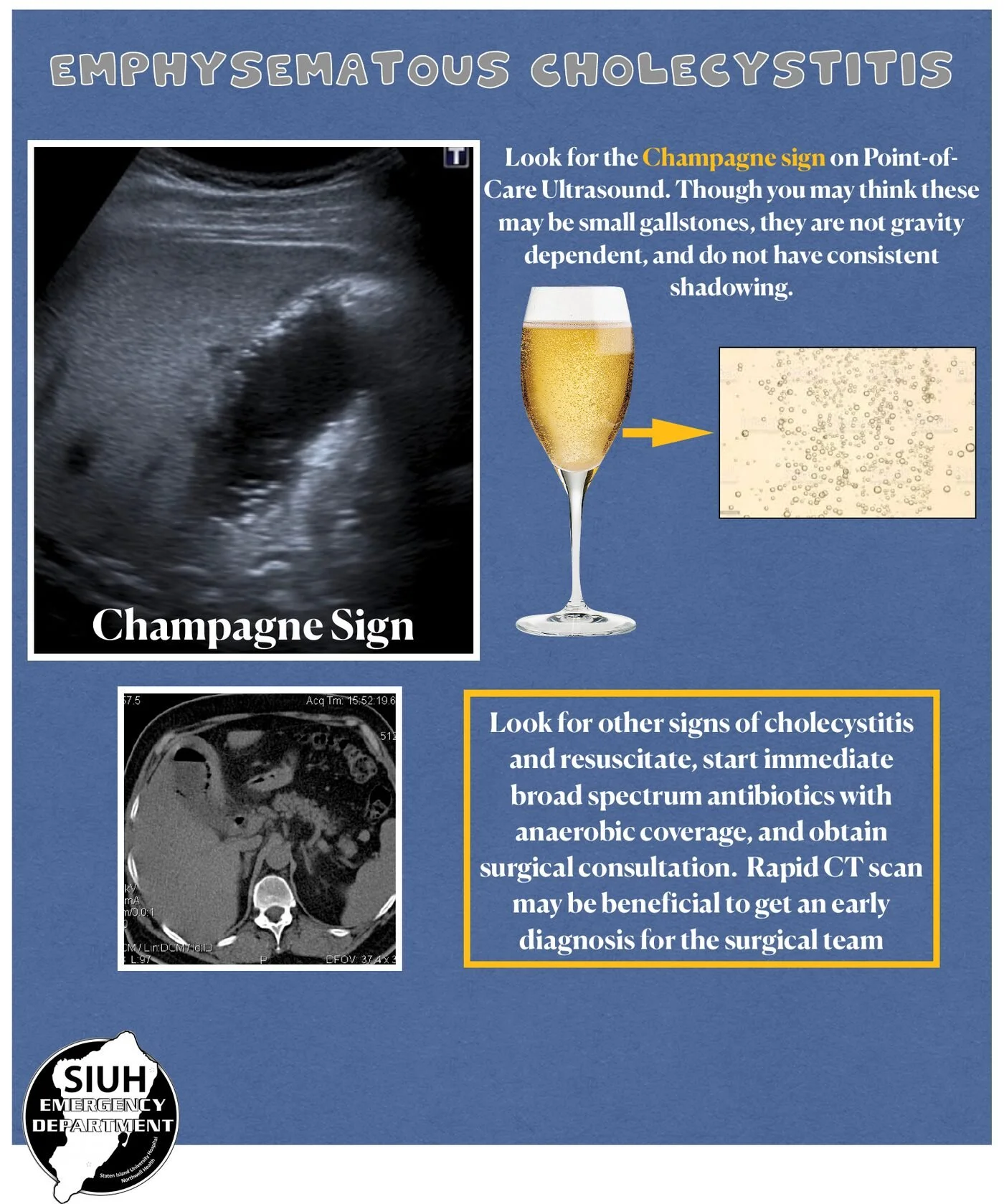
Look for echogenic foci of gas within the gallbladder: the champagne sign on POCUS.
These foci can be mistaken for small gallstones. Unlike stones, they are not gravity dependent and do not cast consistent shadows.
What to do next
Look for other sonographic signs of cholecystitis.
Resuscitate and start immediate broad-spectrum antibiotics with anaerobic coverage.
Obtain surgical consultation.
A rapid CT may help give the surgical team an early diagnosis.
Clinical pearls
Air in the wrong place on POCUS means the patient needs a surgeon now. Do not wait for confirmatory imaging to start antibiotics and call surgery.
Gas produces dirty shadowing and reverberation, not the clean acoustic shadow of a stone.
Non-dependent echogenic foci in the gallbladder should raise concern for emphysematous cholecystitis rather than cholelithiasis.